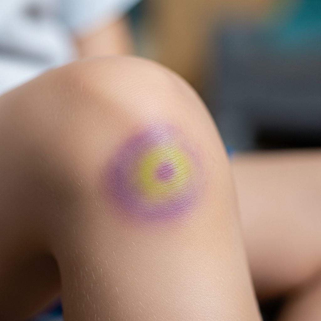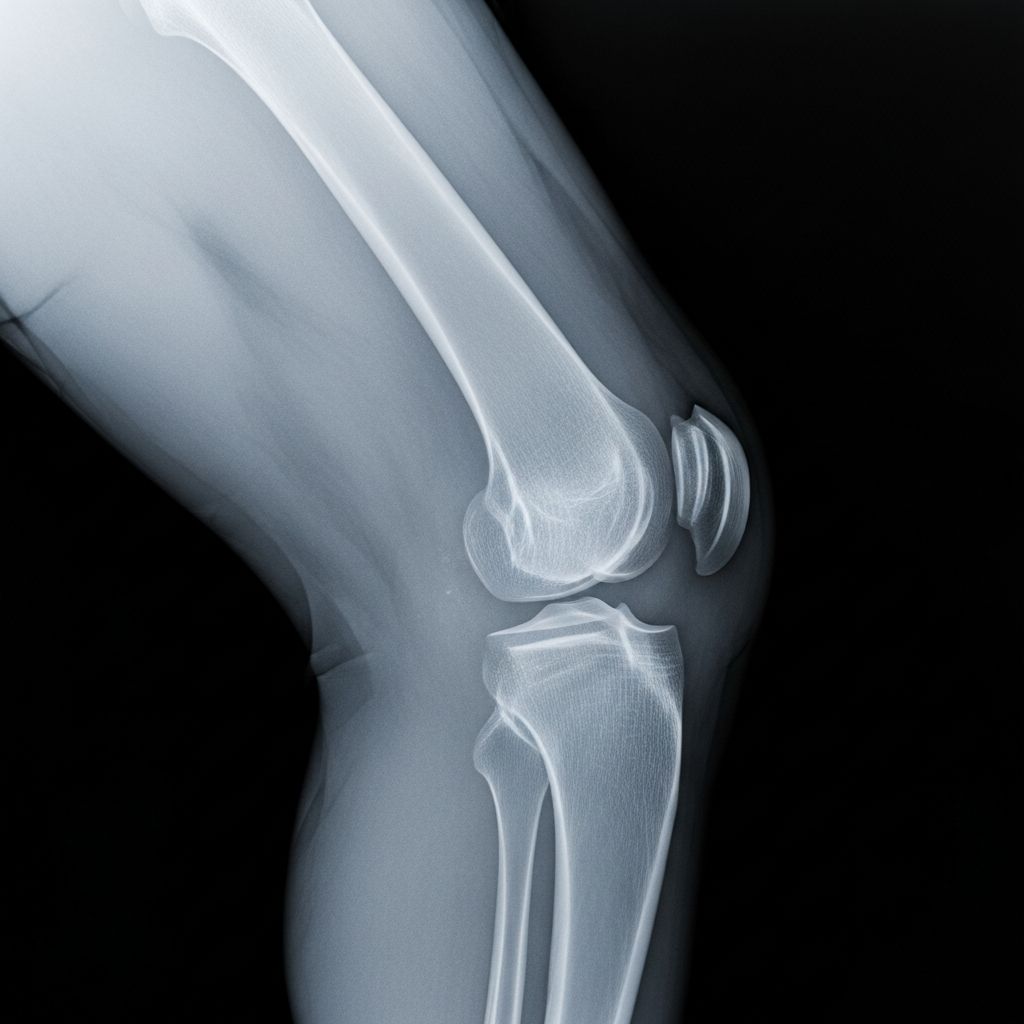
Knee X-rays are a common, quick imaging test used to examine the bones and joints in a child’s knee, helping diagnose injuries, growth issues, or other conditions.
Understanding Knee X-Rays in Pediatric Care
In pediatric medicine, knee X-rays play a crucial role because children’s bones are still developing. These images capture the femur, tibia, fibula, and patella, revealing fractures, alignment problems, or signs of conditions like osteoarthritis in rare early cases. Unlike adults, kids’ growth plates—soft areas of developing bone—appear distinct on X-rays, aiding in the detection of unique pediatric issues such as Osgood-Schlatter disease or avulsion fractures.
The process uses low-dose radiation to produce clear black-and-white pictures of bone structures and some soft tissues. For children, minimizing exposure is key, so technicians use protective shields and precise positioning.
Common Reasons for Knee X-Rays in Kids
Parents often seek knee X-rays when their child experiences persistent pain, swelling, limping, or after a fall or sports injury. These scans help identify:
- Fractures: Breaks in the femur, tibia, fibula, or patella, common in active children.
- Dislocations or misalignments: Such as patella subluxation or tibio-femoral issues.
- Growth plate injuries: Critical in kids, as they can affect future bone development.
- Joint space narrowing: Indicating early cartilage wear or effusion.
- Other issues: Bone cysts, tumors, infections, or post-surgical healing.
In sports like soccer or gymnastics, X-rays detect stress fractures or ligament avulsions, guiding treatment from rest to surgery.
How the Knee X-Ray Procedure Works for Children
The procedure is straightforward and child-friendly, typically lasting 5-10 minutes in a radiology room. A technologist positions the child—standing, sitting, or lying down—for optimal images, often using pillows for comfort.
Standard views include:
- Anteroposterior (AP) view: Front-to-back, assessing alignment and degeneration.
- Lateral view: Side profile, checking patella position and tendon length.
- Sunrise or Merchant’s view: Knee bent, evaluating patellofemoral joint for tilt or tracking issues.
- Optional standing or long-leg views: For alignment in weight-bearing positions.
Children must stay still; fun techniques like games or counting help young ones comply. A lead apron shields the torso, reducing radiation to safe levels.
| View Type | Purpose | Child Positioning |
|---|---|---|
| AP | Overall bone alignment | Standing or supine, knees straight |
| Lateral | Patella and joint depth | Side-lying, knee flexed 30° |
| Sunrise | Patellofemoral tracking | Knee bent 30-45° |
| Standing PA | Functional degeneration | Weight-bearing, knee flexed |
Sometimes, the other knee is imaged for comparison.
Preparing Your Child for a Knee X-Ray
Preparation is minimal. Remove clothing over the knee and any metal items like jewelry. Explain the process simply: “It’s like a quick photo of your knee—no pain, just hold still!” Practice poses at home to ease anxiety.
For toddlers, parental presence and distractions like toys help. No fasting required, and sedation is rare unless the child has severe mobility issues.
Safety and Radiation Concerns for Young Patients
X-rays use ionizing radiation, but doses for knee imaging are very low—less than background radiation from a day’s environment—and safe for children when needed. Benefits outweigh risks for diagnosing serious issues.
Guidelines from bodies like the American College of Radiology emphasize ALARA (As Low As Reasonably Achievable) principles: collimation limits the beam, digital detectors reduce dose, and shields protect gonads and thyroid. No long-term side effects reported from single or occasional knee X-rays.
Interpreting Your Child’s Knee X-Ray Results
A radiologist analyzes images for abnormalities. Key findings include:
- Normal: Clear bones, even joint spaces, proper alignment.
- Fractures: Lines or disruptions in bone continuity.
- Effusion: Fluid buildup darkening suprapatellar area.
- Patella alta: High-riding kneecap on lateral view.
- Tibial plateau issues: Alignment deviations over 0.5 cm.
- Notch width: For ligament sizing in ACL concerns.
Reports use terms like “no acute fracture” or “mild joint narrowing.” Discuss with your pediatrician; follow-ups may include MRI for soft tissues.
When Further Imaging or Treatment Follows
If X-rays show fractures, casting or surgery may be needed. Alignment issues might require orthotics or physical therapy. Growth plate concerns warrant specialist review.
Serial X-rays track healing. For chronic pain, combine with ultrasound or MRI.
FAQs: Knee X-Rays for Kids
Is a knee X-ray painful for children?
No, it’s painless—just positioning and holding still. Mild discomfort from poses is eased with supports.
How long do results take?
Preliminary views immediate; full radiologist report in hours to days, shared at follow-up.
Can X-rays show ligament tears?
Indirectly via avulsions or alignment; MRI better for soft tissues.
Are knee X-rays safe during growth spurts?
Yes, low-dose and targeted; essential for timely diagnosis.
What if my child can’t stay still?
Technologists use distractions; sedation rare, only if medically necessary.
Empowering Parents: Next Steps After X-Ray
Review results with your doctor, ask about implications for school sports or activities. Encourage RICE (Rest, Ice, Compression, Elevation) initially. Nutrition with calcium/vitamin D supports bone health.
Stay proactive: Track symptoms and follow recommendations to ensure your child’s knee recovers fully.
References
- Knee Radiography (X-Rays) — The Knee Doc. Accessed 2026. https://www.thekneedoc.co.uk/investigations/x-rays/
- X-Ray for Osteoarthritis of the Knee — Healthline. 2023-05-15. https://www.healthline.com/health/osteoarthritis/oa-of-the-knee-xray
- Knee X-Ray: Anatomy, Procedure & What to Expect — Cleveland Clinic. 2024-02-20. https://my.clevelandclinic.org/health/diagnostics/23501-knee-x-ray
- Knee X-rays — Don’t Forget the Bubbles. 2023-11-10. https://dontforgetthebubbles.com/knee-x-ray-interpretation/
- X-Ray Exam: Knee — Nemours KidsHealth. 2025-01-01. https://kidshealth.org/en/parents/xray-knee.html