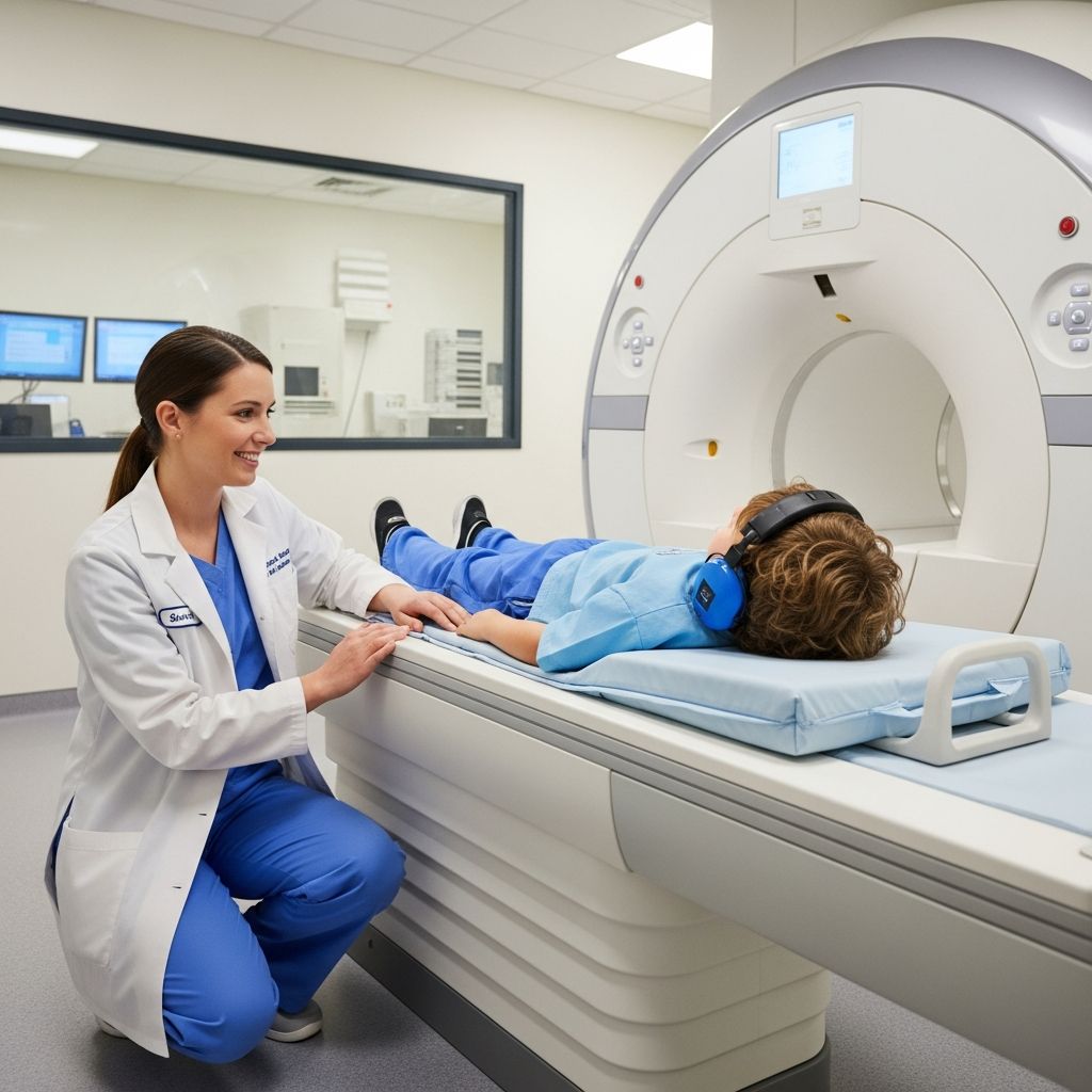
When a doctor recommends a spine MRI for a child, many parents immediately wonder if the test will hurt, whether it is safe, and how to help their child stay calm. This guide explains, in clear language, why spine MRIs are done for children, how the test works, what happens before, during, and after the scan, and how parents can make the experience easier.
What Is a Spine MRI for Children?
MRI stands for magnetic resonance imaging. A spine MRI uses a strong magnet, radio waves, and a computer to create very detailed pictures of the bones, discs, nerves, and soft tissues in a child’s back and neck.
Unlike X-rays or CT scans, MRI does not use ionizing radiation. Instead, it relies on magnetic fields and radiofrequency signals, which are carefully controlled and monitored by trained staff.
Why Doctors Order a Spine MRI
Doctors recommend a spine MRI when they need more information than a physical exam or simpler imaging tests (like X-rays) can provide. The goal is to see detailed images of the spinal cord, nerves, vertebrae, and surrounding tissues to better understand a child’s symptoms.
Common Reasons for a Pediatric Spine MRI
- Persistent back or neck pain that does not improve with rest or basic treatment.
- Leg or arm weakness, numbness, tingling, or changes in walking or balance.
- Suspected spinal curvature problems, such as scoliosis or other abnormal curves.
- Possible spinal cord or nerve compression from a disc, bone, or other structure.
- Injury or trauma to the back or neck that might involve the spinal cord.
- Suspected infection, inflammation, or tumor affecting the spine or spinal cord.
- Follow-up on known spine or nervous system conditions to check progression or response to treatment.
Sometimes a spine MRI is ordered even when symptoms are mild if the doctor is concerned about an underlying condition that needs early diagnosis and monitoring.
How a Spine MRI Works (Kid-Friendly Explanation)
A spine MRI scanner is a large, tube-shaped machine with an opening in the center and a narrow bed that slides in and out. The child lies on the bed while the machine takes pictures.
Inside the scanner, the magnet causes signals in the body’s tissues. Special radio waves and a computer then turn those signals into highly detailed images, slice by slice, allowing doctors to see inside the spine from many angles.
Important Features of MRI Technology
- Strong magnet: Always present, even when the machine seems off, so strict safety rules about metal are followed.
- Radio waves: Used to create signals from body tissues; these are painless and not felt by the child.
- Computer processing: Assembles the data into images that can show even small changes in bones, discs, nerves, and the spinal cord.
Is a Spine MRI Safe for Children?
For most children, a spine MRI is considered very safe. It does not expose the child to radiation, and the magnetic fields and radiofrequency energy are managed by strict safety standards.
The main safety concern is metal. Because the magnet is so powerful, children and anyone entering the room must remove metal objects, and the team carefully screens for implants or devices that might be affected by the magnetic field.
When Extra Safety Precautions Are Needed
- Implanted medical devices (pacemakers, some shunts, certain ear implants).
- Metal fragments in the body, especially near the eyes or brain.
- Certain older surgical clips or hardware that are not MRI-compatible.
The radiology and referring teams review the child’s history and any implanted devices before the scan. In some cases, they may adjust the MRI settings or choose another test.
Preparing Your Child for a Spine MRI
Preparation helps reduce fear and makes it easier for a child to remain still, which is essential for getting clear images. Parents can play a major role by explaining the test in simple, reassuring terms and practicing at home.
What Parents Can Do at Home
- Describe the scanner as a “big camera” that takes pictures of the inside of the back or neck.
- Practice lying very still on the floor or bed while timing your child with a fun game.
- Talk about the loud knocking and humming sounds and compare them to a drum or train.
- Let your child choose a favorite non-metal toy or blanket if the facility allows it.
- Explain that parents can usually stay nearby, depending on the hospital’s rules.
Typical Instructions Before the Appointment
- Arrive early to complete safety forms and answer questions about the child’s medical history.
- Remove all jewelry, hair clips, and clothing with metal snaps or zippers.
- In some cases, follow fasting instructions if sedation or anesthesia is planned.
- Bring a list of medications, allergies, and any implanted devices.
- Share any previous imaging results or surgery records if requested.
What Happens on the Day of the Scan
On the day of the spine MRI, families usually first check in at the imaging center or hospital. After registration, a technologist or nurse will review safety questions, explain the procedure, and answer last-minute concerns.
Step-by-Step MRI Visit
- Check-in and screening: Forms are reviewed, and staff ask about health history, metal implants, and recent illnesses.
- Changing clothes: The child may put on a hospital gown to avoid hidden metal fasteners.
- IV placement (if needed): If contrast dye or sedation is planned, an intravenous line may be placed, usually in the arm or hand.
- Entering the MRI room: Only screened people are allowed, and all metal objects must be left outside.
- Positioning on the table: The child lies on the MRI table; straps, cushions, or special pads help keep them still and comfortable.
- Ear protection: Earplugs or headphones reduce the loud scanner noises.
- Scanning: The table slides into the scanner, and the machine takes a series of picture sets, each lasting a few minutes.
- Completion: Once enough images are taken, the table moves out, and the IV (if used) is removed.
Contrast Dye and Sedation: When Are They Used?
Some spine MRIs are done with a contrast dye, and some children need help remaining still through sedation or anesthesia. Both decisions depend on the child’s age, medical condition, and the type of information the doctor needs.
Contrast Dye
Contrast for MRI is usually a gadolinium-based liquid given through an IV. It makes certain blood vessels, tissues, and inflammatory or abnormal areas more visible, helping doctors distinguish between different structures.
- Often used when there is concern for infection, inflammation, or tumors.
- May be needed for follow-up images during the same visit.
- Side effects are uncommon but can include mild reactions; severe allergic reactions are rare.
Sedation or Anesthesia
Remaining still for the entire scan can be difficult for younger children or those with anxiety, pain, or special needs. In such cases, the medical team may recommend sedation or anesthesia.
- Mild sedation: Medicine helps a child relax or become drowsy but may not fully put them to sleep.
- Deep sedation or anesthesia: The child is more deeply asleep and closely monitored by specialists.
- Parents receive detailed fasting and after-care instructions when sedation is planned.
During the Scan: What Your Child Experiences
Once the scan begins, the child will hear loud tapping, humming, or thumping sounds as the machine changes its magnetic settings to capture different image sequences. Ear protection helps make the noise more tolerable.
The child must lie very still, sometimes holding a particular position. The technologist can usually see and hear the child at all times and may offer reminders and encouragement over an intercom.
Comfort and Coping Strategies
- Some centers allow music or recorded stories through headphones.
- Child-life specialists may use toys, play, or practice sessions to reduce fear.
- Parents may be able to stay in the room, depending on safety rules and their own screening.
- Warm blankets and positioning cushions help children feel secure and less confined.
After the Spine MRI
After the scan, most children can return to normal activities quickly, especially if they did not receive sedation. They can usually eat and drink right away unless told otherwise by the staff.
If sedation or anesthesia was used, nurses monitor the child until fully awake and stable. Slight grogginess, irritability, or unsteadiness can occur but typically improves within a few hours.
When Results Are Available
A specially trained doctor called a radiologist reviews the MRI images, interprets what they show, and sends a report to the referring provider. The child’s doctor then discusses the findings, what they mean, and possible next steps.
Typical Duration and Comfort Levels
A spine MRI usually lasts between 30 and 60 minutes, depending on the area examined (neck, mid-back, lower back, or the entire spine) and whether contrast is used. Extra time may be needed for preparation and recovery.
The test itself is painless. Any discomfort usually comes from holding a still position for a long time, the coldness of the room, or anxiety about the unfamiliar environment.
Risks, Limitations, and Alternatives
While MRI is generally safe, no test is entirely risk-free. Concerns may include rare reactions to contrast dye, complications from sedation or anesthesia, and the possibility that a child cannot complete the scan due to anxiety or movement.
MRI also has limitations. For example, certain types of metal implants may interfere with image quality, and some conditions may still require other tests, such as X-ray, CT scan, or ultrasound, for a complete evaluation.
Spine MRI and Other Imaging Tests
| Test | Uses | Radiation Exposure | Key Strengths |
|---|---|---|---|
| MRI | Detailed view of spinal cord, nerves, discs, and soft tissues. | None (no ionizing radiation). | Excellent soft-tissue contrast; good for complex or unclear problems. |
| X-ray | Overview of bones and alignment; often first test for back issues. | Low dose of ionizing radiation. | Quick, widely available, good for assessing spinal curvature and fractures. |
| CT scan | Detailed bone imaging and some soft-tissue evaluation. | Higher dose of ionizing radiation compared with X-ray. | Very fine detail of bones; useful in trauma or complex bone anatomy. |
| Ultrasound (limited role) | Sometimes used in very young infants for certain spinal issues. | No ionizing radiation. | Portable and radiation-free, but not suitable for all spine questions. |
How Parents Can Advocate for Their Child
Parents are key members of the child’s care team. Asking questions and sharing observations about symptoms, behavior changes, or pain patterns can help the doctor decide whether a spine MRI is necessary and how to plan it safely.
- Ask why the MRI is needed and what information it may reveal.
- Discuss whether contrast or sedation will be used and why.
- Share any history of allergies, reactions to medicines, or previous imaging results.
- Let the team know if the child has sensory sensitivities, anxiety, or special needs.
Frequently Asked Questions (FAQs)
1. Will my child feel anything during the spine MRI?
The scan itself is painless. Your child will not feel the magnetic field or radio waves. Any discomfort usually comes from staying still, lying on the table, or wearing positioning devices that keep the body steady.
2. How long will the whole visit take?
The actual scan is often 30–60 minutes, but plan for extra time for check-in, safety screening, changing clothes, and recovery if sedation is used. In many cases, families spend one to two hours at the imaging facility.
3. Can I stay with my child in the MRI room?
Many centers allow a parent or caregiver to stay in the room as long as they also pass safety screening and remove all metal objects. Policies vary, so ask the facility in advance what to expect.
4. Is sedation always required for children?
No. Older children and some younger ones can complete the scan without sedation, especially with preparation and support. Sedation or anesthesia is more likely for very young children or those who find it hard to stay still or manage anxiety.
5. What should I do if my child is very anxious or claustrophobic?
Share these concerns with the care team before the appointment. They may suggest a mock scanner session, child-life support, relaxation techniques, or mild medication. Explaining the process in advance and practicing stillness at home also helps.
6. Are there long-term risks from a spine MRI?
MRI does not use ionizing radiation, and current evidence supports its safety when appropriate precautions are followed. Long-term problems from the scan itself are considered extremely rare.
7. When will we know the results?
Timing varies by facility, but many reports are available to the referring doctor within a few days. Your child’s doctor will then contact you to explain the findings and discuss any further tests or treatments.
Key Takeaways for Parents
- A spine MRI provides detailed images of a child’s back and neck without using radiation.
- The test is painless, but children must lie very still in a noisy, tube-shaped machine.
- Preparation, clear explanations, and comfort measures greatly reduce anxiety and movement.
- Contrast dye and sedation are used only when needed and with careful monitoring.
- Parents should feel empowered to ask questions and participate in decisions about imaging.