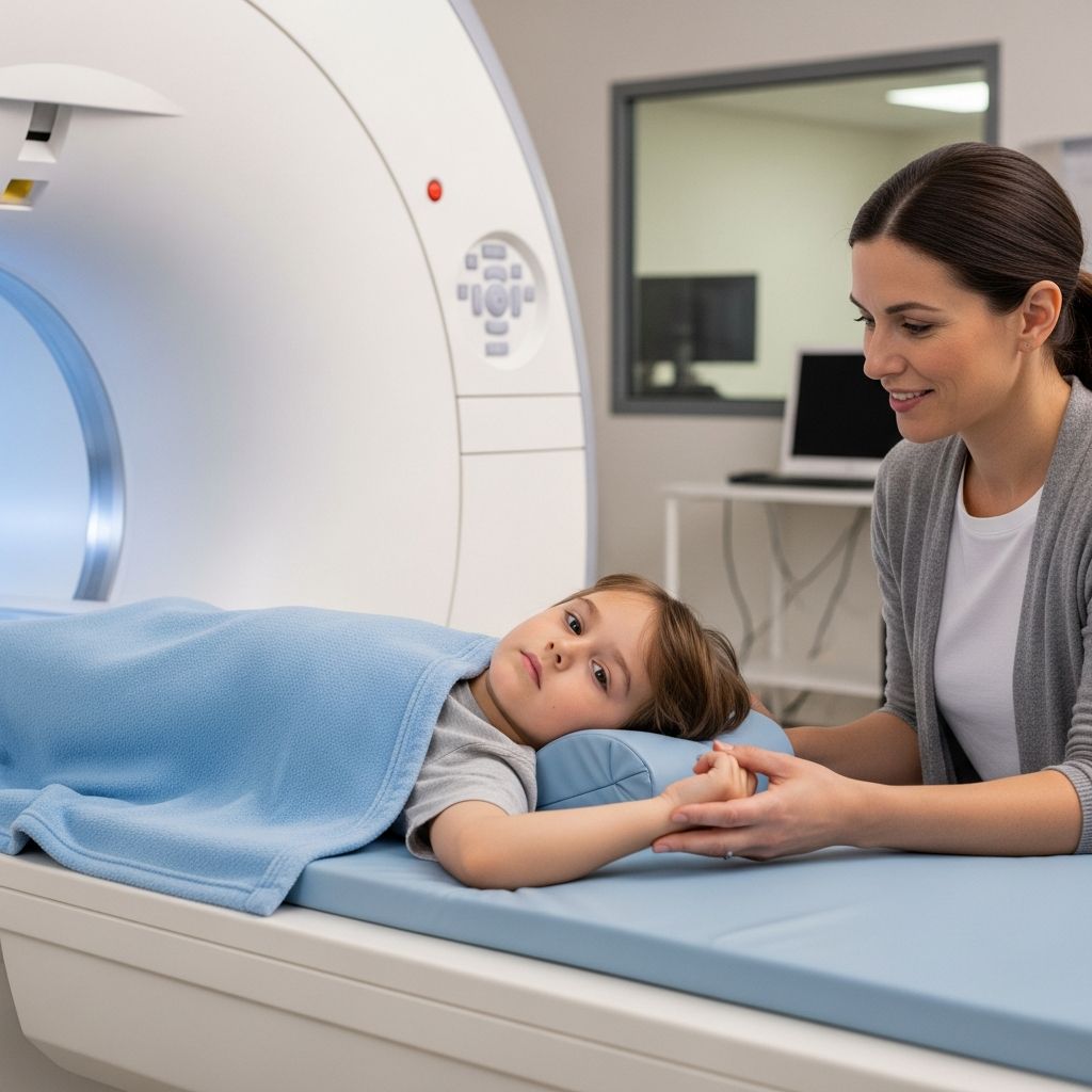
The lumbar spine MRI is a vital diagnostic tool used to examine the lower back in children, providing detailed images of bones, discs, nerves, and soft tissues without invasive procedures. This scan helps identify issues causing pain, weakness, or other symptoms in kids’ lower backs.
Understanding the Lumbar Spine in Children
The lumbar region consists of the five vertebrae (L1 to L5), sacrum, and coccyx, supporting the body’s weight and protecting the spinal cord. In children, this area is still developing, making it susceptible to congenital issues, injuries from sports, or growth-related conditions. Surrounding structures like nerves, ligaments, discs, and blood vessels are crucial for mobility and sensation in the legs.
Unlike adults, children’s spines are more flexible but can experience unique problems such as spondylolisthesis (vertebra slippage), disc herniations from trauma, or inflammatory conditions. Early detection via MRI prevents long-term complications like nerve damage or mobility issues.
Common Reasons for Ordering a Lumbar MRI in Kids
Pediatricians recommend lumbar MRIs when symptoms suggest spinal involvement. Key indicators include:
- Persistent lower back pain unrelieved by rest or medication.
- Leg pain, numbness, tingling, or weakness (sciatica-like symptoms).
- Bladder or bowel control difficulties, signaling nerve compression.
- Fever with back pain, hinting at infection or inflammation.
- Post-injury evaluation for fractures, disc issues, or soft tissue damage.
- Suspected tumors, congenital defects, or conditions like multiple sclerosis.
These scans also monitor treatment progress or pre-surgical planning, ensuring precise interventions.
How Does a Lumbar Spine MRI Work?
MRI technology employs powerful magnets and radio waves to align hydrogen protons in the body, then disrupt and realign them to emit signals. A computer processes these into high-resolution cross-sectional images, excelling at visualizing soft tissues over X-rays or CT scans.
For the lumbar area, sequences capture sagittal (side views), axial (cross-sections), and coronal planes, highlighting vertebrae alignment, disc integrity, nerve roots, and spinal cord health. Contrast agents like gadolinium may enhance visibility of inflammation or tumors if needed.
Preparing Your Child for the MRI Scan
Preparation varies by age and facility protocols. Key steps include:
- Screening for Safety: Complete a questionnaire on metal implants, pacemakers, or claustrophobia. Children with braces may need removal.
- Fasting if Contrast Used: No food/drink 4-6 hours prior for sedation or IV dye.
- Comfort Measures: Explain the process simply; use stories or videos. Bring comfort items like stuffed animals.
- Sedation for Young Kids: Under 6-7 years, mild sedation ensures stillness; pre-procedure check by an anesthesiologist.
- Clothing and Arrival: Wear metal-free clothes; arrive early for paperwork.
Practice lying still with eyes closed to mimic the 30-60 minute scan.
Step-by-Step: What Happens During the Procedure
The process is painless but noisy. Here’s the typical flow:
- Check-In and IV Placement: If contrast is used, an IV line is inserted for dye injection midway.
- Positioning: Child lies supine on a padded table, head stabilized, knees supported with cushions.
- Entering the Scanner: Table slides into the cylindrical magnet; technician communicates via intercom.
- Scanning Sequences: Loud banging sounds as images are taken; headphones or earplugs provided.
- Contrast if Needed: Brief pause for injection; scan continues to capture enhanced images.
- Completion: Table slides out; child rests briefly if sedated.
Parents often stay nearby, visible through a window.
| Factor | Typical Time |
|---|---|
| Without Contrast | 30-45 minutes |
| With Contrast | 45-60 minutes |
| Sedated Child | 60-90 minutes (incl. recovery) |
Ensuring Safety and Minimizing Risks for Children
MRIs are safe, radiation-free, ideal for kids. Rare risks include:
- Allergic Reaction to Contrast: <1% chance; monitored closely.
- Sedation Side Effects: Temporary drowsiness; vital signs watched.
- Noise-Induced Hearing: Mitigated by protection.
- Claustrophobia: Open MRI or sedation helps.
No long-term effects; NSF (nephrogenic systemic fibrosis) is negligible with modern low-dose agents.
Decoding Your Child’s MRI Results
A radiologist analyzes images, reporting to your doctor. Common findings:
- Normal: No action needed.
- Disc Herniation: Bulging/ruptured disc pressing nerves.
- Stenosis: Narrowed canal compressing cord/nerves.
- Inflammation/Infection: Abscesses or discitis.
- Tumors/Cysts: Benign/malignant growths.
- Fractures/Malalignment: Spondylolisthesis or trauma.
Results guide treatments: physical therapy, meds, or surgery.
After the Scan: Recovery and Next Steps
Most kids resume normal activities immediately. Sedated children recover 1-2 hours under observation. Hydrate post-contrast; watch for rash/nausea. Discuss results at follow-up; further tests if unclear.
Comparing MRI to Other Spinal Imaging
| Modality | Pros | Cons | Best For |
|---|---|---|---|
| MRI | Detailed soft tissue; no radiation | Expensive; time-consuming | Nerves, discs, tumors |
| CT | Fast; bone detail | Radiation exposure | Fractures |
| X-ray | Quick; cheap | Limited soft tissue | Alignment |
MRI is gold standard for comprehensive lumbar evaluation in children.
Frequently Asked Questions (FAQs)
Is lumbar MRI safe for young children?
Yes, it’s non-invasive and radiation-free. Sedation ensures cooperation without risks for most kids.
How long do results take?
Preliminary same day; full report 1-3 days.
Can my child eat before the scan?
Yes, unless contrast or sedation requires fasting.
What if my child has metal in their body?
Screening determines eligibility; some implants are MRI-conditional.
Does the scan hurt?
No pain, just loud noises and need to stay still.
Empowering Parents: Questions to Ask Your Doctor
- Why is this specific scan needed for my child?
- Are there alternatives with less preparation?
- What sedation options are available?
- How will results impact treatment?
- Any immediate concerns post-scan?
Active involvement ensures optimal care.
References
- What Does a Lumbar Spine MRI Show Your Doctor? — Charlotte Radiology. 2023. https://go.charlotteradiology.com/blog/what-does-a-lumbar-spine-mri-show-your-doctor/
- Lumbar MRI Scan: Purpose, Procedure, and Risks — Healthline. 2023-05-10. https://www.healthline.com/health/lumbar-mri-scan
- Lumbar MRI Scan: What to expect and results — Medical News Today. 2023-04-27. https://www.medicalnewstoday.com/articles/323693
- Magnetic Resonance Imaging (MRI): Lumbar Spine — KidsHealth (Nemours). 2024. https://kidshealth.org/en/parents/mri-lumbar.html
- Making sense of MRI of the lumbar spine — RACGP. 2012-11-01. https://www.racgp.org.au/afp/2012/november/making-sense-of-mri-of-the-lumbar-spine
- What to Know About Your Lumbar Spine MRI — DocPanel. 2023. https://docpanel.com/what-know-about-your-lumbar-spine-mri
- Lumbar spine MRI scan, protocols, positioning and planning — MRIMaster (YouTube). 2017-10-20. https://www.youtube.com/watch?v=XQKysY5VEi8
- MRI of the Lumbar Spine: About This Test — MyHealth Alberta. 2023. https://myhealth.alberta.ca/Health/aftercareinformation/pages/conditions.aspx?hwid=abq5157