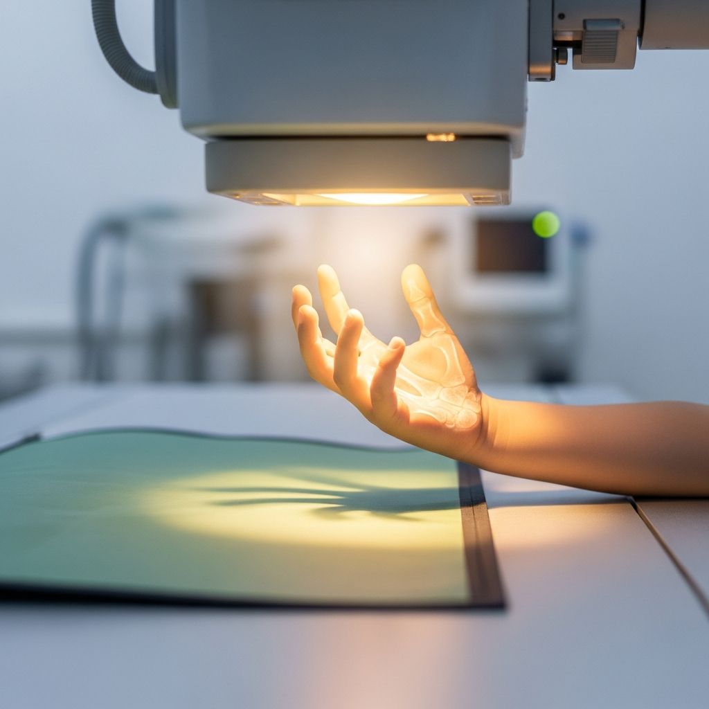
Understanding Hand and Finger Imaging in Pediatric Care
Hand and finger X-rays represent one of the most common and straightforward imaging procedures performed on children. These diagnostic tools allow healthcare providers to visualize the bones, joints, and surrounding tissues of the hand without requiring invasive procedures or sedation. The accessibility and speed of hand X-rays make them invaluable in pediatric medicine, helping doctors identify fractures, assess growth patterns, evaluate joint conditions, and determine overall skeletal development. Understanding what this procedure involves helps parents prepare their children appropriately and alleviates unnecessary anxiety about the imaging process.
Why Pediatricians Order Hand and Finger X-Rays
Healthcare providers request hand and finger X-rays for several important diagnostic reasons. One of the most common applications involves assessing a child’s bone age, which refers to the skeletal maturity level compared to chronological age. This measurement helps pediatricians determine whether a child is growing at an appropriate rate or if growth abnormalities might indicate underlying health conditions requiring intervention. By comparing the appearance and development of bones in the hand to established standards for various ages, doctors can identify growth disorders early and adjust treatment plans accordingly.
Hand X-rays are essential for evaluating suspected fractures and injuries. When a child reports pain, swelling, or visible deformity in the fingers, hand, or wrist following a fall or accident, an X-ray provides definitive confirmation of bone breaks and helps determine the severity and location of the injury. This information guides treatment decisions, from simple rest and splinting to more complex interventions requiring specialist consultation.
Additionally, these imaging studies help doctors evaluate various medical conditions affecting hand structure and function, including joint disorders, bone infections, developmental anomalies, and inflammatory conditions. The hand’s relatively simple anatomy and accessibility make it an ideal location for imaging practice and training.
How the Hand X-Ray Procedure Functions
Hand X-rays operate using electromagnetic radiation to create detailed images of internal bone structures. The X-ray machine directs a carefully controlled beam of radiation through the hand, which passes through soft tissues at varying rates depending on density. Bones, being denser than surrounding soft tissues, absorb more radiation and appear white on the resulting image, while softer tissues appear in shades of gray.
The procedure involves minimal radiation exposure. The actual time the X-ray beam activates typically lasts less than one second, delivering an extremely small dose of radiation comparable to natural background radiation exposure over several days. Modern digital X-ray technology has further reduced radiation doses while improving image quality, making the procedure safer than ever before.
What Parents and Children Should Expect
Before the appointment, parents should dress their child in comfortable, loose-fitting clothing that allows easy access to the hand and arm. Most facilities ask children to remove any metal jewelry, watches, or bracelets, as metal objects can create artifacts that obscure the image quality. Arriving a few minutes early allows time for check-in and explanation of the procedure by the radiology technologist.
During the examination, the child will typically sit at the X-ray table and place their hand flat on a special platform or against the imaging plate. The technologist will position the hand to capture the necessary views, usually requiring images from at least two different angles. The child must remain completely still for 2-3 seconds while each image is captured, as any movement during radiation exposure results in blurred pictures that require retakes.
The technologist may ask the child to hold their breath briefly or adjust hand position between images. Older children generally understand these instructions well, while younger children benefit from simple, reassuring explanations about what they’ll experience. The entire procedure, from positioning through image verification, typically takes only 5 to 15 minutes.
Comfort Considerations During the Procedure
Most children experience no discomfort during hand X-rays. The radiation itself is completely imperceptible—children cannot feel, see, taste, or hear X-rays as they pass through their body. However, some children report mild discomfort from the cool temperature in the X-ray room or from the hardness of the imaging table. The positioning required for optimal images occasionally feels awkward, particularly if the child has an existing hand or arm injury causing pain.
Parents often accompany their children into the X-ray room, providing reassurance and emotional support. When parents remain in the room, they typically wear a lead apron to protect their reproductive organs from radiation exposure, though the small radiation dose to bystanders is negligible. The presence of a familiar adult significantly reduces anxiety in young children.
Safety Profile and Radiation Exposure
Hand and finger X-rays are among the safest imaging procedures available in pediatric medicine. The radiation dose delivered during a hand X-ray is extremely small—so minimal that the benefit of obtaining an accurate diagnosis far outweighs any theoretical risk from radiation exposure. To put this in perspective, a child receives more radiation from natural background sources in a single day than from a typical hand X-ray procedure.
Modern pediatric facilities specifically calibrate X-ray machines for children, using the smallest radiation dose necessary to produce diagnostic quality images. This child-specific approach differs from adult X-ray settings and ensures that imaging benefits are maximized while radiation exposure is minimized. Parents concerned about radiation exposure can discuss these safety measures with their healthcare provider, who can explain the specific clinical reasons why the imaging is necessary and beneficial.
Bone Age Assessment: A Common Application
One specialized use of hand X-rays involves determining a child’s skeletal or bone age. This assessment compares the appearance of bones and growth plates in the hand to standardized reference images for different ages. Bone age differs from chronological age in children with growth disorders, hormonal imbalances, or developmental delays.
The radiologist carefully examines the size, shape, and degree of ossification (hardening) of various hand and wrist bones. Growth plates—the areas of developing tissue near the ends of bones—show characteristic changes as children mature. By comparing these features to established standards, radiologists can estimate skeletal age with reasonable accuracy. This information helps pediatricians determine whether a child’s growth rate is appropriate or whether further evaluation and treatment might be warranted.
Preparing Your Child for the Procedure
Preparation significantly improves the imaging experience for anxious children. Parents should explain to their child in simple, age-appropriate language what will happen during the X-ray, emphasizing that:
- The procedure is painless and very fast
- The child will sit comfortably at a table with their hand positioned flat
- The machine will take pictures of their hand bones
- They need to stay very still for just a few seconds
- A parent can stay in the room with them
- The technologist is experienced with children and wants to help them feel comfortable
Reading age-appropriate books about medical procedures or watching simple videos about X-ray procedures can further reduce anxiety. Some facilities employ child life specialists who explain procedures in developmentally appropriate ways and help children cope with medical situations more effectively.
After the X-Ray Examination
Once imaging is complete, children can immediately return to normal activities with no restrictions or recovery period needed. Unlike procedures requiring sedation, hand X-rays involve no medication and no side effects. Parents should expect radiologist results within 48 to 72 business hours, though some facilities provide preliminary results more quickly. The radiologist examines the images and generates a detailed report that the child’s physician reviews before discussing findings and treatment recommendations with the family.
Comparing Hand X-Rays to Other Imaging Options
Hand X-rays are the first-line imaging study for most hand and finger concerns because they are quick, accessible, inexpensive, and effective for evaluating bone structures. Advanced imaging modalities like ultrasound, MRI, or CT scans are occasionally needed for specific situations involving soft tissue evaluation or complex fracture patterns, but these procedures are more time-consuming and expensive than standard X-rays.
Ultrasound imaging requires no radiation and can visualize soft tissues like tendons and ligaments effectively, but it cannot evaluate bone detail as clearly as X-rays. MRI provides excellent soft tissue detail and three-dimensional information but requires longer scan times and may need sedation in uncooperative young children. CT scans deliver higher radiation doses than conventional X-rays. For most pediatric hand and finger evaluations, conventional X-rays provide the ideal balance of diagnostic accuracy, safety, speed, and cost-effectiveness.
Frequently Asked Questions About Hand and Finger X-Rays
Will my child feel pain during the hand X-ray?
No. The X-ray procedure itself is painless, and children cannot feel the radiation passing through their body. If a child has an existing hand injury, they may experience mild discomfort from positioning the injured area, but the radiologist will work to position it as comfortably as possible to obtain necessary images.
How much radiation does a hand X-ray expose my child to?
Hand X-rays deliver minimal radiation exposure—less than a second of actual beam activation and an extremely small dose. The radiation exposure is comparable to natural background radiation received over several days and is far outweighed by the diagnostic benefits of identifying fractures or growth abnormalities.
Can I stay with my child during the X-ray?
Yes. Most facilities allow parents to remain in the X-ray room with their child for reassurance and support. If you stay in the room, you’ll be asked to wear a protective lead apron to shield your reproductive organs, though radiation exposure to bystanders is negligible.
How long does a hand X-ray take?
A typical hand X-ray examination takes 5 to 15 minutes from start to finish, including positioning, image capture, and verification. The actual radiation exposure time is less than one second.
What does bone age tell us about my child’s growth?
Bone age assessment compares your child’s skeletal maturity to age-matched standards. If bone age is significantly different from chronological age, it may indicate growth disorders, hormonal imbalances, or developmental delays that warrant further evaluation and possible treatment.
Will my child need to remove their clothing for the X-ray?
No. Children remain fully clothed during hand X-rays. They should wear comfortable, loose-fitting clothing that allows easy access to the hand and arm, and should remove jewelry or metal bracelets that might interfere with image quality.
How soon will I get the X-ray results?
Radiologists typically review X-ray images and prepare reports within 48 to 72 business hours. Your child’s physician will review the radiologist’s report and discuss findings and recommendations with you.
Is it necessary to prepare my child before the X-ray appointment?
While no medical preparation is needed, explaining the procedure to your child in simple, age-appropriate language can reduce anxiety. Describing the procedure as fast and painless, and mentioning that you’ll stay nearby, helps most children feel more comfortable.
References
- Diagnostic Testing: Bone Age X-Ray — University of Florida Powell Center for Rare Diseases. https://raredisease.powellcenter.med.ufl.edu/pediatric-care-center/diagnostic-testing/
- Pediatric X-ray Exam — Radiologyinfo.org. https://www.radiologyinfo.org/en/info/pediatric-xray
- X-Ray Exam: Finger (for Parents) — Kids Health. https://kidshealth.org/HumanaKentucky/en/parents/xray-finger.html
- X-Ray Imaging Tests — Children’s Hospital of Philadelphia. https://www.chop.edu/treatments/general-radiography-x-ray
- Hand X-Ray: Anatomy, Procedure & What To Expect — Cleveland Clinic. https://my.clevelandclinic.org/health/diagnostics/23518-hand-x-ray