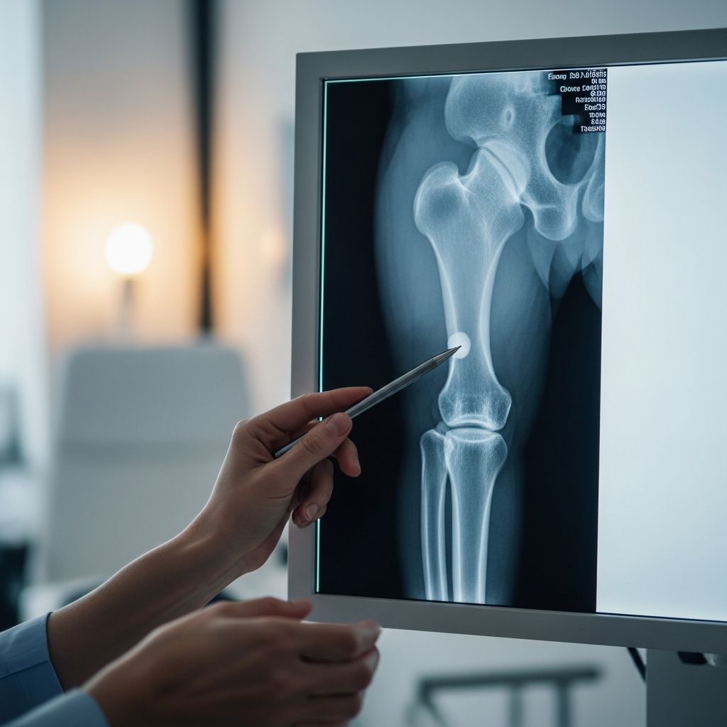
The femur, or thigh bone, is the longest and strongest bone in the human body, connecting the hip to the knee. In children, X-rays of this bone are common diagnostic tools used to assess injuries, growth issues, or congenital conditions. This guide provides parents with detailed, actionable information on when a femur X-ray is necessary, how to ready your child, procedural details, and post-exam care.
Understanding the Role of Femur X-Rays in Pediatric Care
Femur X-rays help visualize the bone’s structure, identifying fractures, misalignments, or abnormalities. They are quick, non-invasive, and vital for timely treatment in active kids prone to falls or sports injuries. Unlike MRI or CT scans, X-rays use low radiation doses, making them suitable for children.
These imaging studies capture the proximal (hip-end), shaft, and distal (knee-end) femur portions. Multiple views ensure comprehensive coverage due to the bone’s length.
Common Reasons for Ordering a Femur X-Ray in Kids
- Trauma and Fractures: Falls, sports accidents, or car crashes often cause femur breaks. X-rays confirm if the bone is intact or fractured, showing break type and location.
- Growth and Development Checks: Monitors bone lengthening, alignment, or conditions like Blount’s disease.
- Infections or Tumors: Detects bone infections (osteomyelitis) or rare tumors by revealing density changes.
- Joint Evaluations: Assesses hip or knee involvement when femur issues suspected.
- Follow-Up Imaging: Tracks healing after prior injuries or surgeries.
Pediatric orthopedists rely on these images for precise diagnoses, as clinical exams alone may miss subtle issues.
Preparing Your Child for a Femur X-Ray
Preparation is straightforward, focusing on comfort and cooperation. Explain the process simply: “It’s like a quick photo of your leg bones—no needles or pain.” Practice staying still with fun timers.
| Age Group | Preparation Tips | Potential Challenges |
|---|---|---|
| Infants/Toddlers | Bring a favorite toy or blanket; feed beforehand to avoid fussiness. | May need gentle restraint; parental holding allowed. |
| School-Age (5-12) | Describe superhero poses; watch kid-friendly videos. | Fear of machines; reassure with tech’s presence. |
| Teens | Discuss privacy; involve in positioning decisions. | Embarrassment; explain medical necessity. |
Remove metal objects like jewelry or belts from the leg area. No fasting required unless sedation planned for very young or anxious children.
Step-by-Step: What Happens During the Procedure
The exam typically takes 15-30 minutes in a radiology suite. Child lies supine on the table; tech positions the leg for optimal views.
- AP Proximal Femur: Leg internally rotated 15-20 degrees; captures hip to mid-thigh using 14×17 inch cassette.
- AP Distal Femur: Knee in true AP; overlaps prior image to cover full shaft down to knee.
- Frog-Leg Lateral (Proximal): Knee flexed 45 degrees, leg abducted like a frog; profiles femoral neck.
- Lateral Distal: Affected side down; knee flexed, captures knee up to mid-femur.
For trauma cases unable to move, cross-table laterals use horizontal beams. Techs use lead shields for pelvis/gonads, minimizing exposure.
Patient must hold still 2-3 seconds per image; young kids may need parental assistance or mild sedation.
Radiation Safety and Risks for Pediatric Patients
X-rays emit ionizing radiation, but pediatric doses are minimized (e.g., 0.001-0.01 mSv per femur series, comparable to background radiation). ALARA principle (As Low As Reasonably Achievable) guides protocols: low kVp (75-80), adjusted mAs.
- Shields protect non-imaged areas.
- Digital detectors reduce repeat exposures.
- Benefits outweigh risks for necessary diagnostics.
Pregnant mothers should inform staff; alternatives like ultrasound considered for hips.
Interpreting Your Child’s Femur X-Ray Results
Radiologists analyze for:
- Fractures: Lines, displacements (transverse, spiral).
- Alignment: Proper neck-shaft angle (135-145 degrees in kids).
- Density: Uniform trabeculae; abnormalities signal pathology.
- Joints: Hip/knee spaces symmetric.
Normal: Clear cortex, no overlaps. Reports detail findings, recommend follow-up. Orthopedists explain: greenstick fractures common in kids due to flexible bones.
After the X-Ray: Recovery and Next Steps
No recovery needed; resume activities immediately unless injury restricts. Results available same day; digital sharing speeds consultations.
Discuss with doctor: immobilization (casts), surgery (intramedullary nails for severe cases), or physical therapy.
Special Considerations for Young Children and Infants
Infants tolerate positioning well but need swaddling. Portable units for bedridden kids. Growth plates (physes) appear as dark lines—normal but monitored to avoid premature closure.
FAQs for Parents on Pediatric Femur X-Rays
Is a femur X-ray painful for my child?
No, it’s painless. Discomfort only from holding positions if injured.
How many X-rays are taken?
Usually 4 views for full coverage due to femur length.
Can I stay with my child?
Yes, wearing lead apron behind barriers.
What if my child can’t stay still?
Sedation or immobilization aids used; rare for older kids.
Are there alternatives to X-rays?
Ultrasound for soft tissues; MRI for complex cases, but X-ray first-line for bones.
How soon do results come?
Preliminary in minutes; full report within hours.
Preventing Femur Injuries in Active Children
Promote safety: helmets for biking, proper sports gear, home fall-proofing. Nutrition (calcium, vitamin D) supports bone health. Regular check-ups catch issues early.
In summary, femur X-rays empower quick, accurate care for kids’ thigh bone concerns, balancing diagnostics with safety.
References
- Femur x-ray protocol — YouTube (Radiographic Procedures). Accessed 2026. https://www.youtube.com/watch?v=2Su4Lg8PibQ
- Radiographic Positioning of the Femur and Tib Fib — CE4RT.com. Accessed 2026. https://ce4rt.com/rad-tech-talk/resources/radiographic-positioning-of-the-femur-and-tib-fib/
- Femur Shaft Fractures (Broken Thighbone) — American Academy of Orthopaedic Surgeons (OrthoInfo). 2023-01-01. https://orthoinfo.aaos.org/en/diseases–conditions/femur-shaft-fractures-broken-thighbone/
- Pelvis X-ray: Purpose, Procedure & Risks — Cleveland Clinic. 2023-05-15. https://my.clevelandclinic.org/health/diagnostics/23519-pelvis-x-ray
- X-Ray Exam: Upper Leg (Femur) — Rady Children’s Health. Accessed 2026. https://www.rchsd.org/health-article/x-ray-exam-upper-leg-femur/