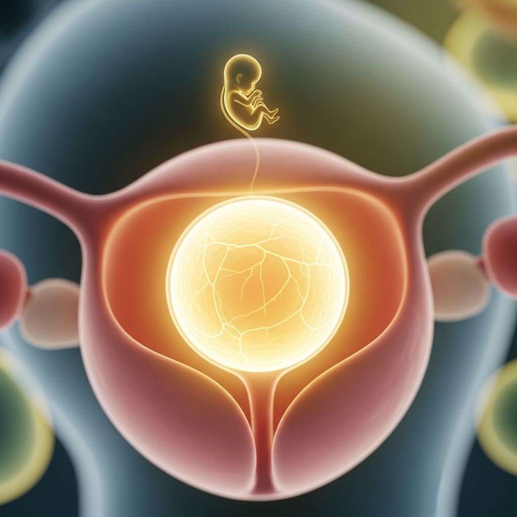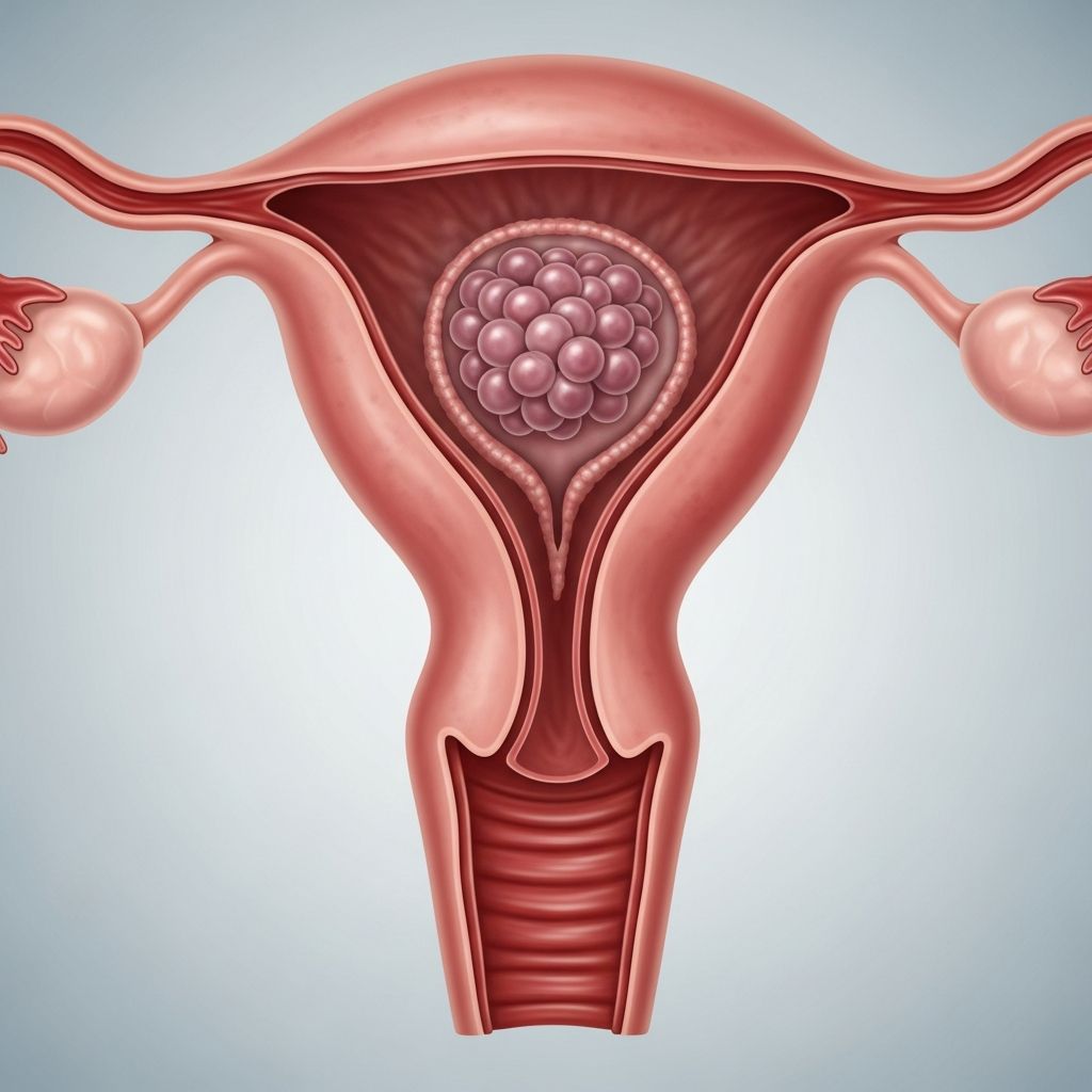
Molar pregnancy represents a rare yet significant deviation from normal gestation, where placental tissue proliferates abnormally within the uterus, preventing fetal development. This condition, part of gestational trophoblastic disease (GTD), affects roughly 1 in 1,000 pregnancies and demands prompt medical attention to avert severe complications.
Defining the Condition and Its Core Characteristics
In typical pregnancies, the fertilized egg implants and develops into an embryo supported by a nourishing placenta. Conversely, a molar pregnancy features the growth of a hydatidiform mole—a cluster of fluid-filled sacs resembling grapes—instead of viable fetal structures. This anomaly arises from fertilization errors, leading to trophoblast cells, which normally form the placenta, expanding uncontrollably.
The absence of a functional embryo distinguishes molar pregnancies, resulting in inevitable pregnancy loss if undiagnosed. Early detection through routine prenatal checks is crucial, as untreated cases can progress to persistent trophoblastic disease or rare malignant forms like choriocarcinoma.
Primary Types of Molar Pregnancies
Molar pregnancies classify into two distinct categories based on genetic composition and clinical presentation:
- Complete Molar Pregnancy: No maternal chromosomes contribute; an empty egg is fertilized by one or two sperm, duplicating paternal DNA to yield 46 chromosomes all from the father. No embryo or normal placental tissue forms—only swollen villi.
- Partial Molar Pregnancy: The egg retains maternal chromosomes, but fertilization by two sperm introduces excess paternal sets, totaling 69 chromosomes. Minimal fetal tissue may develop but is non-viable, accompanied by abnormal placental growth.
Complete moles occur more frequently and pose higher risks for malignant transformation compared to partial ones, which sometimes mimic miscarriages.
Genetic Underpinnings and Origin Mechanisms
At conception, human cells require 46 chromosomes—23 from each parent—for balanced development. Molar pregnancies disrupt this harmony through aberrant fertilization. In complete moles, maternal genetic material is lost or inactivated, with paternal chromosomes duplicating via androgenesis. Partial moles stem from dispermy, where twin sperm fertilize one egg, overloading paternal genetics.
Though exact triggers remain elusive, these errors highlight vulnerabilities in gamete fusion. Research underscores that such chromosomal imbalances halt embryonic progression, redirecting cellular energy toward trophoblastic overgrowth.
Who Faces Elevated Risks?
Certain demographics experience heightened susceptibility:
| Risk Factor | Details | Increased Odds |
|---|---|---|
| Maternal Age | Under 20 or over 40, peaking after 43 | Up to 5-10 times higher |
| Prior Molar Pregnancy | History of one elevates recurrence | 1-2% chance |
| Nutritional Deficiencies | Low intake of protein, folate, carotene | Associated in some studies |
| Geographic Variation | Higher in Asia, Latin America | Up to 1 in 120 pregnancies |
Women with these profiles should prioritize early ultrasounds and hCG monitoring. Extremes of reproductive age correlate with ovum quality issues, amplifying genetic mishaps.
Recognizing Warning Signs Early On
Symptoms often emerge by the first trimester, mimicking routine pregnancy discomforts but escalating rapidly:
- Profuse vaginal bleeding, sometimes with grape-like tissue passage
- Uterine size exceeding gestational age expectations
- Severe nausea and vomiting beyond typical morning sickness
- Preeclampsia before 20 weeks—high blood pressure, proteinuria
- Hyperthyroidism symptoms: rapid heartbeat, anxiety
- Ovarian cysts causing pelvic pain
- Anemia from blood loss
These red flags, especially bleeding in early pregnancy, warrant immediate obstetric evaluation. Elevated hCG levels, often exceeding 100,000 mIU/mL, further signal abnormality.
Diagnostic Approaches and Confirmation Methods
Diagnosis integrates clinical assessment, imaging, and lab tests:
- Ultrasound: Primary tool revealing ‘snowstorm’ pattern in complete moles or irregular cysts in partial ones; absent fetal heartbeat confirms.
- hCG Quantification: Markedly high serum levels diverge from normal pregnancy curves.
- Histopathology: Post-evacuation tissue analysis verifies trophoblastic proliferation and genetic makeup.
Color Doppler enhances ultrasound by detecting absent vascularity in molar tissue. Serial hCG tracking post-treatment monitors for persistence.
Standard Treatment Protocols
Surgical evacuation via dilation and curettage (D&C) under anesthesia removes molar tissue promptly. Suction curettage minimizes bleeding risks, followed by sharp curettage for completeness. Hysterectomy serves extreme cases in older patients not desiring future pregnancies.
Rh-negative women receive RhoGAM to prevent isoimmunization. Post-procedure, strict hCG surveillance—weekly until undetectable, then monthly for six months—guards against GTD. Contraception is advised during this period to avoid confounding new pregnancies.
Potential Complications and Vigilance Needs
While most resolve post-evacuation, 15-20% of complete moles develop persistent GTD, necessitating chemotherapy like methotrexate for invasive or metastatic disease. Hyperthyroidism and theca lutein cysts typically regress spontaneously.
Thyroid function stabilizes with mole removal, but monitoring averts crises. Rare choriocarcinoma demands multi-agent regimens with high cure rates over 90%.
Journey to Recovery and Emotional Healing
Physical recovery spans weeks, with light activities resuming soon after D&C. hCG normalization signals success, permitting conception after 6-12 months. Emotional support addresses grief akin to miscarriage, through counseling or groups.
Future pregnancies carry low recurrence risk with vigilant monitoring. Nutritional optimization—folate-rich diets—may mitigate odds.
Frequently Asked Questions About Molar Pregnancy
Is molar pregnancy preventable?
No definitive prevention exists, but prenatal care, age awareness, and nutrition support risk reduction.
Can a molar pregnancy produce a healthy baby?
No; by definition, it lacks viable fetal development in complete cases, and partial ones result in non-survivable anomalies.
How long does hCG monitoring last after treatment?
Typically 6-12 months, with contraception until levels normalize and stabilize.
What diet helps after a molar pregnancy?
Focus on folate (leafy greens, legumes), protein, and carotenes (carrots, sweet potatoes) for overall reproductive health.
Does molar pregnancy increase cancer risk?
Low overall; 1-5% for persistent GTD, treatable with excellent prognosis if caught early.
Steps for Future Family Planning
Post-molar patients should schedule preconception counseling. Early ultrasounds at 6-8 weeks detect anomalies swiftly. Genetic counseling benefits high-risk individuals. Most achieve successful term pregnancies thereafter, underscoring resilience.
References
- Molar pregnancy – Symptoms and causes — Mayo Clinic. 2023-10-15. https://www.mayoclinic.org/diseases-conditions/molar-pregnancy/symptoms-causes/syc-20375175
- Molar pregnancy — March of Dimes. 2024-05-01. https://www.marchofdimes.org/find-support/topics/miscarriage-loss-grief/molar-pregnancy
- Hydatidiform Mole — StatPearls, NCBI Bookshelf. 2023-07-17. https://www.ncbi.nlm.nih.gov/books/NBK459155/
- Molar Pregnancy: Symptoms, Causes, & Treatments — WebMD. 2024-02-20. https://www.webmd.com/baby/what-is-molar-pregnancy
- Understanding Molar Pregnancy — Natera. 2023-11-10. https://www.natera.com/resource-library/anora/understanding-molar-pregnancy/