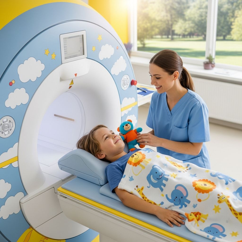
When a child has ongoing lower back or leg problems, a lumbar spine MRI can give doctors detailed images of the bones, nerves, and soft tissues in the lower back so they can understand what is going on and plan the best treatment.
Understanding the Lumbar Spine
The lumbar spine is the lower part of the backbone, just above the hips. It supports much of the body’s weight and protects important nerves that travel from the brain down into the legs.
This area includes:
- Five strong lower back bones called lumbar vertebrae.
- Soft pads between the bones, known as disks, which work like shock absorbers.
- Nerves that branch out from the spinal cord and help control movement and sensation in the legs.
- Muscles, ligaments, and other tissues that help the spine bend, twist, and stay stable.
What Is a Lumbar Spine MRI?
A lumbar spine MRI (magnetic resonance imaging) is a painless imaging test that uses a powerful magnet and radio waves to create clear, detailed pictures of the lower back. Unlike X-rays and CT scans, it does not use ionizing radiation.
During the scan, the machine takes multiple image slices of the spine, which a computer combines into views that help doctors see bones, disks, nerves, and surrounding tissues in great detail.
Why a Doctor Might Order This Test
Doctors recommend a lumbar spine MRI when symptoms suggest a problem in the lower back that cannot be fully explained by a physical exam or simpler tests. It is especially useful when symptoms last a long time or are severe.
Common reasons a doctor may order this test for a child include:
- Persistent lower back pain that does not improve with rest or basic treatment.
- Pain that travels from the lower back into the buttocks or legs.
- Numbness, tingling, or weakness in one or both legs.
- Changes in walking, balance, or coordination.
- Concerns about bladder or bowel control, such as leaking or trouble going.
- Possible injury to the spine from a fall, sports impact, or accident.
- Suspicion of infection, inflammation, or growths in or around the spine.
Conditions an MRI Can Help Detect
A lumbar spine MRI does not diagnose everything, but it can provide important clues. Based on these images, doctors can look for several types of problems in the lower back.
| Area | Possible Issue | What It May Cause |
|---|---|---|
| Disks | Bulging, tears, or herniation | Back pain, leg pain, tingling, or weakness if nerves are pressed |
| Bones (vertebrae) | Fractures, structural changes, or abnormal alignment | Pain, posture changes, or nerve compression |
| Spinal canal | Narrowing (stenosis) or crowding of nerve structures | Leg pain, weakness, or problems walking |
| Soft tissues | Inflammation, infection, or masses | Pain, stiffness, or other general symptoms |
| Nerve roots | Compression, irritation, or swelling | Shooting pain, numbness, or reduced strength |
How Safe Is a Lumbar Spine MRI?
MRI is considered a very safe test because it does not involve radiation, but it does use a strong magnet, so safety screening is essential. Children usually tolerate the test well when they know what to expect and feel supported.
Important safety points include:
- Any metal in or on the body must be checked before the scan.
- Certain implants, devices, or metal fragments may make MRI unsafe or require special planning.
- Staff will ask detailed questions about surgeries, implants, and injuries to ensure the scan is appropriate.
When Sedation or Anesthesia May Be Used
Staying very still is essential for clear MRI images. For many children, especially younger ones, this can be challenging, so sedation (medicine to make a child drowsy) or anesthesia (medicine to make a child sleep) may be recommended.
The decision to use these medicines depends on:
- The child’s age and ability to follow instructions.
- The length and complexity of the scan.
- How anxious or uncomfortable the child feels about the procedure.
Preparing Your Child for the Scan
Good preparation can lower fear and help the scan go more smoothly. Parents play a key role in explaining the test in a calm, age-appropriate way.
Helpful preparation steps include:
- Describing the MRI as a big camera that uses magnets to take pictures of the back.
- Explaining that the machine makes loud knocking or thumping sounds, but they are normal.
- Letting your child know that they must lie very still, like a statue, for a short time.
- Practicing “still time” at home by timing how long your child can remain in one position.
- Bringing a comfort item if allowed, such as a favorite blanket or small stuffed toy without metal.
Clothing and Belongings
Because of the magnet, metal objects are not allowed in the scanner room. This includes jewelry, coins, keys, hair clips, and some clothing items with zippers or snaps.
On the day of the scan, your child may be asked to:
- Change into a hospital gown free of metal parts.
- Remove glasses, watches, jewelry, and removable dental devices.
- Leave electronic items, toys with batteries, and cards with magnetic strips outside the room.
What Happens on the Day of the MRI
Knowing the basic steps of the visit can help both you and your child feel more at ease. Although each hospital or imaging center has its own routine, the general process is similar.
Check-In and Screening
When you arrive, you will sign in and complete safety forms that ask about your child’s medical history, implants, and possible metal exposure. Staff will answer questions and explain what will happen next.
If sedation or anesthesia is planned, additional checks such as vital signs, recent illnesses, and fasting times are usually reviewed.
Entering the MRI Area
After screening, you and your child are brought to the MRI suite. In many centers, a parent can stay in the room during the scan, as long as safety rules are followed and no metal items are carried in.
The MRI machine looks like a large tube with an opening in the middle and a table that slides in and out. The spine images are taken while your child lies on this table.
Step-by-Step: During the Scan
The actual scan involves several short imaging sequences. Each sequence may take a few minutes, and your child will hear loud repeating sounds while images are being taken.
A typical sequence of events may include:
- Your child lies on the table, usually on the back, and is positioned so the lower spine is in the center of the scanner.
- Soft pads, straps, or cushions may be used to help your child stay comfortable and still.
- Special equipment called a coil is placed around or under the lower back to improve image quality.
- Ear protection, such as earplugs or headphones, is provided to soften the loud sounds.
- The table slides into the opening of the MRI machine until the lower back is in the correct position.
- The technologist operates the scanner from a nearby room and communicates through an intercom.
- Your child hears noises like tapping, banging, or humming during image collection; these sounds are normal.
- Between sequences, there may be brief pauses when the machine is quieter and your child can relax, but must remain still.
How Long Does It Take?
The time in the MRI room varies, but most lumbar spine scans take between 20 and 45 minutes of actual imaging. Extra time may be needed for preparation, positioning, and if sedation or anesthesia is used.
The main factor that can extend the exam is movement, because blurred pictures sometimes must be repeated. Remaining as still as possible helps keep the scan shorter and more comfortable.
After the MRI: What to Expect
Once the images are taken, the table slides out of the machine and the technologist removes any straps or coils. If your child did not receive sedation or anesthesia, they can usually stand up and go home shortly after.
If sedating medicines were used, your child will spend extra time in a recovery area until fully awake and stable. Staff will monitor breathing, heart rate, and comfort level before discharge.
Recovery Tips After Sedation or Anesthesia
If your child was given medicines to help with sleep or relaxation, mild side effects are possible as the medicines wear off.
Common post-scan care steps include:
- Keeping your child supervised for the rest of the day, as they may still feel tired or unsteady.
- Offering light foods and clear liquids first, then returning to normal meals if tolerated.
- Following any activity or school restrictions provided by the medical team.
Who Looks at the Images?
After the test, a doctor specially trained in reading medical images, called a radiologist, studies the pictures. The radiologist examines the bones, disks, spinal canal, and surrounding tissues for signs of injury, structural changes, or disease.
The radiologist then writes a report for the doctor who ordered the scan, describing any significant findings and how they may relate to your child’s symptoms.
How and When You Get Results
Results are usually not given immediately in the imaging department. Instead, the ordering doctor reviews the radiologist’s report and explains the findings at a follow-up visit or phone call.
During the discussion, you may learn:
- Whether the scan looked normal or showed any concerning changes.
- How the findings might explain your child’s pain or other symptoms.
- What additional tests or treatments, if any, are recommended.
Comparing Lumbar MRI With Other Tests
Doctors sometimes choose between different imaging methods depending on the question they are trying to answer. Each type has strengths and limitations.
| Test | Main Strengths | Limitations |
|---|---|---|
| Lumbar spine MRI | Excellent detail of soft tissues, disks, nerves, and spinal cord; no radiation | Loud sounds, enclosed space, more sensitive to movement, may require sedation in children |
| X-ray | Quick, widely available, good for viewing bones and alignment | Limited view of disks and nerves; uses a small amount of radiation |
| CT scan | Good bone detail, faster than MRI | Uses more radiation than X-ray; less detail of soft tissues than MRI |
| Ultrasound | No radiation, portable, useful for some soft tissues | Not typically used for detailed images of the lumbar spine itself |
Helping Your Child Feel More Comfortable
An MRI can feel intimidating to a child, but there are many ways to make the experience more positive. Simple coping strategies can reduce fear and improve cooperation.
Ideas that often help include:
- Using simple, honest language and avoiding scary or confusing terms.
- Encouraging your child to ask questions and express feelings about the test.
- Practicing slow breathing or counting games to stay calm during the scan.
- Asking if music, stories, or child-life specialists are available to provide distraction.
Questions to Ask the Care Team
As a parent, being informed helps you advocate for your child and feel more confident in the decisions being made. Before and after the scan, consider asking questions such as:
- Why is a lumbar spine MRI the best test for my child right now?
- Will my child need sedation or anesthesia for this exam, and what are the risks?
- How long will the appointment take from start to finish?
- Can I stay in the room with my child during the scan?
- When and how will we receive the results?
- How will the results influence my child’s treatment plan?
Frequently Asked Questions (FAQs)
Is a lumbar spine MRI painful?
No, the scan itself does not cause pain. Your child will need to lie still on a table, which may be uncomfortable if they already have back pain, but staff can help with pillows or supports.
Will my child be alone in the MRI machine?
Your child will be inside the scanner, but not alone. The technologist can see, hear, and talk with your child the entire time, and many centers allow a parent to remain in the room if safety rules are followed.
What if my child is scared of tight spaces?
If your child is anxious about enclosed spaces, let the staff know ahead of time. They can offer extra support, explain what to expect, and discuss options such as mild sedation if appropriate.
Are there risks from the magnet?
The main concern with the magnet is its effect on metal objects and certain medical implants. Careful screening is done before the scan to identify any items that could be affected so that the exam can proceed safely or be adjusted if needed.
What if the MRI shows something abnormal?
An abnormal result does not always mean a serious condition, but it does mean that the doctor has found changes that might explain your child’s symptoms. Your child’s doctor will discuss what the findings mean and suggest next steps such as observation, physical therapy, medication, or further testing.
Key Takeaways for Parents
- A lumbar spine MRI creates detailed pictures of the lower back without using radiation.
- Doctors use this test to better understand ongoing back or leg problems, especially when symptoms are persistent or complex.
- Good preparation, open communication, and supportive staff help children handle the scan with less fear and more confidence.