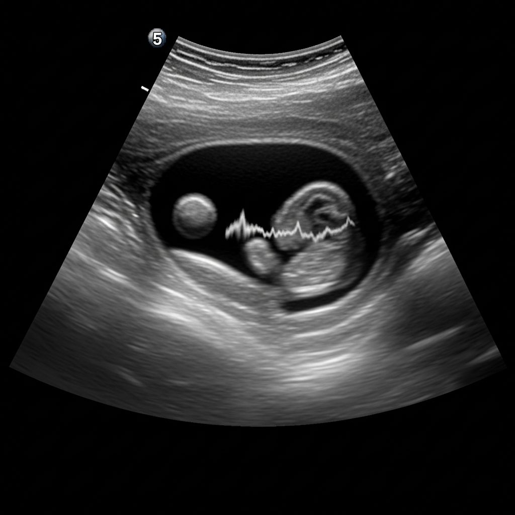
The fetal pole represents one of the earliest visible signs of embryonic development during pregnancy ultrasounds, typically emerging around 5.5 to 6 weeks after the last menstrual period. This small structure, often located adjacent to the yolk sac within the gestational sac, serves as a critical marker for assessing pregnancy progression and viability.
Earliest Signs of Pregnancy on Ultrasound
Ultrasound imaging in the initial weeks of pregnancy reveals progressive milestones. At approximately 4 to 5 weeks, the gestational sac—a fluid-filled structure in the uterus—becomes the first detectable feature, signaling implantation of the fertilized egg.
By 5 to 5.5 weeks, the yolk sac appears as a small, round bubble inside the gestational sac, providing initial nourishment to the embryo before the placenta fully develops.
- Gestational sac: Visible at hCG levels of 1,000-2,000 mIU/mL via transvaginal ultrasound.
- Yolk sac: Measures 3-5 mm, essential for early nutrient transfer.
These structures set the stage for the fetal pole’s emergence, confirming an intrauterine pregnancy and ruling out some ectopic risks early on.
What Exactly Is the Fetal Pole?
The fetal pole is the initial visualization of the embryo itself, appearing as a thickened, bright (hyperechoic) area next to the yolk sac. It measures just 1-2 mm at first detection and grows rapidly, reaching up to 30 mm by 10 weeks.
This pole structure curves slightly, with the head end distinguishable from the tail-like lower portion, which later forms the body’s backbone.
Key functions include:
- Enabling crown-rump length (CRL) measurements for precise gestational dating.
- Indicating embryonic viability when paired with cardiac activity.
- Helping differentiate normal development from anomalies like blighted ovum.
| Gestational Age | Expected Findings | Typical Size |
|---|---|---|
| 5.5-6 weeks | Fetal pole visible | 2-4 mm CRL |
| 6-7 weeks | Heartbeat detectable | 5+ mm CRL |
| Up to 10 weeks | Rapid growth | Up to 30 mm |
Such measurements guide due date estimates, especially when menstrual cycles are irregular.
Timing and Detection Methods
Transvaginal ultrasound is the gold standard for early detection due to its proximity to the uterus, often spotting the fetal pole at 5.5 weeks, while abdominal scans may lag by 1-2 weeks.
Development timeline:
- Weeks 4-5: Gestational sac only.
- Weeks 5-6: Yolk sac and fetal pole.
- Weeks 6-7: Cardiac flicker via M-mode.
hCG levels correlate with visibility: Gestational sac at 1,000-2,000 mIU/mL; fetal pole soon after.
Heartbeat Detection: A Key Milestone
Once the fetal pole reaches 5 mm CRL, a heartbeat—visible as a fluttering motion—is typically detectable around 6 weeks.
This pulsing confirms viability, with rates starting at 90-110 beats per minute, increasing to 140-170 by 9 weeks.
Absence of heartbeat at ≥5-6 mm CRL or ≥16-18 mm gestational sac without pole prompts further evaluation for miscarriage or anembryonic gestation.
Interpreting an Absent Fetal Pole
Not seeing the fetal pole at 6 weeks isn’t always alarming; it may simply mean it’s too early, especially if dates are uncertain.
Follow-up scans in 7-10 days are common. Persistent absence with a large sac (>16-18 mm) suggests blighted ovum, a non-viable pregnancy lacking an embryo.
- Possible causes: Inaccurate dating, recent miscarriage, or ectopic.
- Next steps: Serial hCG tests and repeat ultrasound.
Bedside ultrasound aids quick assessment but pairs with quantitative beta-hCG for accuracy.
Potential Complications and Ectopic Pregnancy
An ectopic pregnancy occurs when the gestational sac implants outside the uterus, often in fallopian tubes—a medical emergency.
Ultrasound signs include absent intrauterine sac or fetal pole in abnormal locations. Pseudo-gestational sacs appear in 10-20% of ectopics, necessitating caution.
Other concerns:
- Blighted ovum: Sac without embryo.
- Miscarriage risk if no heartbeat by expected time.
- Multiples: Multiple sacs/poles in twin pregnancies.
Preparing for Your Early Ultrasound
Expect a full bladder for abdominal scans or none for transvaginal. Results provide reassurance or guide interventions.
Discuss with your provider: Last menstrual period, symptoms, hCG trends. Access reports promptly via patient portals for review.
Advances in Early Pregnancy Imaging
Modern ultrasound offers 3D/4D views later, but 2D transvaginal excels early. AI-assisted measurements improve precision in CRL and heartbeat detection, enhancing outcomes.
Guidelines from bodies like ACOG emphasize timing: Viability scans at 7-8 weeks if bleeding or history of loss.
Frequently Asked Questions
What if no fetal pole at 6 weeks?
It could be too early; repeat in a week. If sac >18 mm without pole, non-viable pregnancy likely.
When is heartbeat visible?
Around 6 weeks when CRL ≥5 mm.
Transvaginal vs. abdominal ultrasound?
Transvaginal detects earlier (5.5 weeks vs. 7-8 weeks).
Does fetal pole confirm twins?
Multiple poles indicate multiples.
hCG level for fetal pole visibility?
Typically after 2,000-3,600 mIU/mL.
References
- Patience is key: Understanding the timing of early ultrasounds — UT Southwestern Medical Center. 2023-05-15. https://utswmed.org/medblog/patience-key-understanding-timing-early-ultrasounds/
- Fetal pole – Global Ultrasound Institute — Global Ultrasound Institute. 2024-02-10. https://globalultrasoundinstitute.com/pocuspedia/fetal-pole/
- Early Pregnancy Ultrasound: What to Expect at Different Weeks — Roswell OBGYN. 2023-11-20. https://roswellobgyn.org/blog/early-pregnancy-ultrasound-what-to-expect-at-different-weeks/
- Early Fetal Development — American Pregnancy Association. 2024-01-05. https://americanpregnancy.org/healthy-pregnancy/pregnancy-health-wellness/early-fetal-development/
- Week 6 of Pregnancy — London Pregnancy Clinic. 2023-09-12. https://www.londonpregnancy.com/week-6-of-pregnancy/
- Fetal Pole: Ultrasound, Anatomy & Function — Cleveland Clinic. 2024-08-22. https://my.clevelandclinic.org/health/body/22546-fetal-pole
- 6-Week Pregnancy Ultrasound — PocketHealth. 2024-03-18. https://www.pockethealth.com/patient-resources/6-week-pregnancy-ultrasound/
- Early Pregnancy | Sonoguide — American College of Emergency Physicians (ACEP). 2023-07-30. https://www.acep.org/sonoguide/basic/early-pregnancy