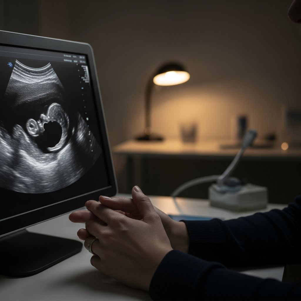
Detecting a fetal heartbeat at exactly 8 weeks isn’t always straightforward due to the embryo’s small size and developmental stage. While cardiac activity often begins around 5-6 weeks, reliable visualization via ultrasound typically requires waiting a bit longer for clearer results.
Understanding Fetal Heart Development in Early Pregnancy
The journey of a baby’s heart starts early. By the fifth week after conception, primitive cardiac tissue forms and begins pulsing, though it’s more like electrical activity in a tubular structure than a fully formed heart. This activity strengthens over the following weeks, transitioning into a recognizable heartbeat detectable by medical imaging.
At 8 weeks gestational age, the embryo measures about the size of a raspberry, roughly 1-2 centimeters from crown to rump. The heart is beating at 110-160 beats per minute, but its tiny scale and the mother’s abdominal layers can obscure detection with certain methods.
- Week 5-6: Initial cardiac pulsing detectable via transvaginal ultrasound in some cases.
- Week 7-8: Heartbeat visible on transvaginal scans for most viable pregnancies, but abdominal scans may miss it.
- Week 9-10: More consistent detection across ultrasound types as the embryo grows.
These timelines vary based on exact conception date, equipment quality, and technician expertise. Gestational age is calculated from the last menstrual period, which can sometimes overestimate or underestimate true embryonic age by several days.
Why You Might Not Detect a Heartbeat at 8 Weeks
Several factors contribute to absent heartbeat detection at this stage, most of which are benign. Miscalculated dates are common; if ovulation occurred later, the embryo might be younger than assumed, lacking sufficient development for visualization.
Home devices like fetal Dopplers add another layer of challenge. Manufacturers claim detection from 8 weeks, but professionals advise waiting until 12-16 weeks. The uterus remains pelvic until late first trimester, positioning the embryo too deep for sound waves to penetrate effectively.
| Method | Earliest Reliable Detection | Challenges at 8 Weeks |
|---|---|---|
| Transvaginal Ultrasound | 6-7 weeks | Possible but not guaranteed; depends on growth. |
| Abdominal Ultrasound | 8-10 weeks | Often too early; embryo too small. |
| Home Doppler | 12-16 weeks | Rarely effective before 12 weeks due to positioning. |
| Stethoscope | 20+ weeks | Not feasible in first trimester. |
Anatomic variations, such as fibroids or excess adipose tissue, can further hinder signals. Even in healthy pregnancies, scans at 6-8 weeks may show only a gestational sac and yolk sac without a visible heartbeat, resolving on follow-up.
Types of Ultrasounds and What to Expect
Early pregnancy ultrasounds fall into two main categories: transvaginal and transabdominal. Transvaginal probes insert near the cervix for proximity to the uterus, ideal for 8-week checks. They reveal the gestational sac, yolk sac, fetal pole, and often a flickering heartbeat.
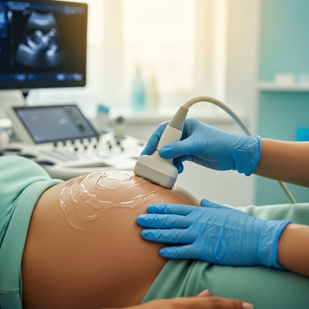
Transabdominal scans apply gel to the belly and use an external probe. At 8 weeks, they confirm pregnancy location and due date but may not show the heartbeat clearly until later.
During the appointment:
- Confirmation of intrauterine pregnancy (rules out ectopic).
- Measurement of crown-rump length for dating accuracy.
- Heartbeat assessment via real-time imaging.
If no heartbeat appears, providers schedule a repeat scan in 7-10 days. This interval allows growth, turning potential concerns into confirmations of viability.
The Role of At-Home Fetal Monitoring Devices
Consumer fetal Dopplers tempt expectant parents with portability, but limitations abound in early pregnancy. The device’s sound waves struggle against the pelvic uterus, often picking up maternal pulse instead.
Normal fetal heart rates range 110-160 bpm, accelerating with movement. Early use risks false negatives, heightening anxiety without benefit. Experts recommend professional ultrasounds over home attempts before 12 weeks.
To use safely later:
- Apply ultrasound gel to the probe.
- Position below navel, near pubic bone.
- Move slowly in circular motions.
- Follow device instructions precisely.
Interpreting Heart Rate Patterns and Normal Ranges
A reassuring heartbeat shows steady rhythm within 110-160 bpm. Variability of 5-25 bpm indicates healthy autonomic function. Deviations prompt further evaluation, like nonstress tests from 32 weeks or fetal echocardiograms for arrhythmias.
Signs warranting attention:
- Persistent rate below 110 bpm.
- Rate exceeding 160 bpm without movement.
- Irregular patterns unresponsive to activity.
Red Flags: When Absence of Heartbeat Signals Concern
While often normal, no heartbeat with a fetal pole measuring over 7mm or after two scans spaced a week apart raises miscarriage risk. Blighted ovum (empty sac) or embryonic demise may present similarly. Immediate follow-up clarifies viability.
Risk factors include advanced maternal age, prior losses, or bleeding/cramping. Always report symptoms promptly for timely intervention.
Steps to Take If No Heartbeat Is Detected
Stay calm—most cases resolve positively. Contact your provider for rescheduling. Track symptoms like spotting or pain, and prepare questions:
- What’s my exact gestational age?
- When is the follow-up scan?
- Any precautions meanwhile?
Lifestyle supports viability: rest, hydration, prenatal vitamins, and avoid stressors. Emotional support via partners, forums, or counseling aids coping.
Emotional Impact and Coping Strategies
Waiting induces anxiety, amplified by online anecdotes. Reframe uncertainty as opportunity for growth confirmation. Journaling, mindfulness, or support groups foster resilience during limbo periods.
Professional mental health resources benefit high-anxiety pregnancies, ensuring holistic care.
Frequently Asked Questions
Can you always see a heartbeat at 8 weeks?
No, not always. Transvaginal ultrasounds detect it in most viable pregnancies by 7-8 weeks, but dating errors or equipment limits may delay visualization until 9 weeks.
Is it safe to use a home Doppler at 8 weeks?
Not recommended. Professional sonographers avoid it before 13 weeks due to inaccuracy; wait until second trimester for reliable home use.
What if no heartbeat on follow-up scan?
Outcomes vary. Many progress normally with time, but persistent absence may indicate loss. Discuss options like expectant management or intervention with your doctor.
When does the fetal heart fully form?
Basic activity starts week 5; four-chamber structure develops by week 9-10, with full maturation continuing post-birth.
How accurate are early ultrasounds for due dates?
Highly accurate in first trimester, within 5-7 days, outperforming later scans.
References
- Baby Fetal Doppler Monitor: Listening to Baby’s Heartbeat — V6CO. Accessed 2026. https://v6co.com/baby-fetal-doppler-monitor-listening-to-babys-heartbeat
- What Can I See On an 8-Week Ultrasound? — Healthline. 2023-10-15. https://www.healthline.com/health/pregnancy/8-week-ultrasound
- Pregnancy Fetal Heart Monitoring — WebMD. 2024-05-20. https://www.webmd.com/baby/pregnancy-fetal-heart-monitoring
- Ultrasound Scans — Miscarriage Association. 2025-01-10. https://www.miscarriageassociation.org.uk/information/worried-about-pregnancy-loss/ultrasound-scans/