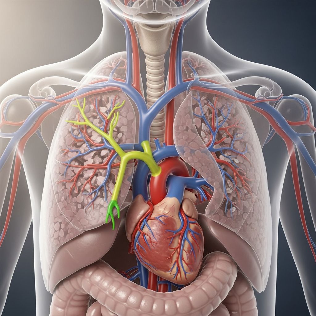
The aorta is the body’s largest artery and the main “highway” that carries oxygen-rich blood from the heart to every organ and tissue. When the curved portion of the aorta, called the aortic arch, develops differently before birth, it can lead to a group of conditions known as aortic arch anomalies. These differences may affect how blood flows or how nearby structures such as the windpipe (trachea) and food pipe (esophagus) function.
This guide explains what aortic arch anomalies are, how they can affect children, how doctors diagnose and treat them, and what families can expect over time.
1. The Aortic Arch: A Quick Overview
To understand aortic arch anomalies, it helps to know the basics of normal aortic anatomy.
- Aorta: the main artery that transports oxygen-rich blood from the left ventricle of the heart out to the body.
- Aortic arch: the curved segment of the aorta that connects the ascending aorta (coming up from the heart) to the descending aorta (traveling down through the chest and abdomen).
- Major branches of the arch:
- Brachiocephalic (innominate) artery – usually the first and largest branch, feeding the right arm and right side of the head and neck.
- Left common carotid artery – supplies blood to the left side of the brain and head.
- Left subclavian artery – delivers blood to the left arm.
The aortic arch bends over the heart like a gentle curve in a river. From there, blood flows through its branches to the brain and arms, and through the descending aorta to the chest and abdomen.
2. What Are Aortic Arch Anomalies?
Aortic arch anomalies are structural differences in the way the aorta and its branches form before birth. They are a type of congenital heart defect, meaning they are present from birth.
In these conditions, the arch may:
- Curve or travel on the right side instead of the usual left side.
- Form a complete or partial ring around the trachea and esophagus (called a vascular ring).
- Have branches that come off in an unusual order or from unexpected locations.
- Be narrowed (coarctation) or interrupted (a gap in continuity).
Some children have only an aortic arch anomaly; others may have additional heart differences, such as a hole between heart chambers or valve problems.
3. Common Types of Aortic Arch Anomalies
Doctors describe aortic arch anomalies based on the path of the arch and how the branches are arranged.
| Type of Anomaly | Key Feature | Possible Effect |
|---|---|---|
| Right aortic arch | Arch passes to the right side of the trachea instead of the left. | May be harmless or part of a vascular ring causing airway/esophagus pressure. |
| Double aortic arch | Two arches (right and left) form a complete ring around trachea and esophagus. | Often significant breathing and feeding problems in infancy due to compression. |
| Aberrant subclavian artery | One arm artery comes off in an unusual position, sometimes behind the esophagus. | May cause swallowing problems if it presses on the esophagus. |
| Coarctation of the aorta | Segment of the arch or descending aorta is abnormally narrow. | Can lead to high blood pressure in the upper body and poor blood flow to the lower body. |
| Interrupted aortic arch | Complete break in continuity between ascending and descending aorta. | Severe, life-threatening condition in newborns if not treated quickly. |
4. Why Do These Anomalies Happen?
During early pregnancy, a complex set of paired embryonic arteries called the pharyngeal (aortic) arches remodel into the single aortic arch seen after birth. The normal adult left-sided arch is mainly formed from the left fourth aortic arch and parts of neighboring segments.
When this remodeling process changes course—because certain segments persist when they usually regress, or regress when they usually persist—an aortic arch anomaly can result.
For many families, no clear cause is found. Contributing factors can include:
- Genetic changes or chromosomal conditions (for example, some right aortic arches and conotruncal heart defects are associated with 22q11.2 deletion).
- Other heart defects that develop alongside the arch anomaly.
- In rare cases, family history of congenital heart disease.
Most parents did nothing to cause an aortic arch anomaly. These conditions typically result from early developmental events that are beyond anyone’s control.
5. How Aortic Arch Anomalies Affect Children
The impact of an aortic arch anomaly depends on its type and severity.
5.1 Effects on Blood Flow
- Normal or near-normal flow: Some right aortic arches and mild branching differences may not significantly change blood flow and can remain asymptomatic.
- Obstruction to flow:
- Coarctation narrows the vessel, making the heart work harder to push blood through the tight segment.
- Interrupted arch prevents direct flow from the heart to the lower body, relying on temporary fetal connections after birth.
- Uneven pressure: Narrowing in the arch can cause high blood pressure in the arms and head, with weaker pulses in the legs.
5.2 Effects on Breathing and Swallowing
When the arch and its branches form a vascular ring, they can squeeze the trachea and esophagus. This may cause:
- Noisy or high-pitched breathing (stridor)
- Chronic cough or wheezing that does not respond well to asthma medications
- Difficulty feeding or swallowing
- Choking, gagging, or frequent vomiting with feeds
- Slow weight gain in infants
6. Signs and Symptoms Parents Might Notice
Symptoms can appear anytime from the newborn period to later childhood. Some children are diagnosed only after imaging is done for another reason.
6.1 Possible Symptoms in Babies
- Fast or labored breathing
- Noisy breathing, especially when feeding or crying
- Poor feeding or tiring quickly with feeds
- Blue color around the lips or face during episodes
- Not gaining weight as expected
6.2 Possible Symptoms in Older Children
- Chronic cough or recurrent respiratory infections
- Difficulty swallowing solid foods
- Chest pain or discomfort with activity
- Headaches or nosebleeds (in coarctation with high blood pressure)
- Decreased exercise tolerance compared with peers
Some aortic arch anomalies cause few or no symptoms and may be found incidentally during heart imaging done for another concern.
7. How Aortic Arch Anomalies Are Diagnosed
Diagnosis usually involves several steps and tests guided by a pediatric cardiologist.
7.1 Medical History and Physical Exam
- Review of symptoms such as breathing difficulties, feeding problems, or poor growth.
- Listening to the heart and lungs for murmurs or abnormal breath sounds.
- Checking pulses and blood pressures in arms and legs to look for differences suggestive of coarctation.
7.2 Imaging Tests
- Echocardiogram (heart ultrasound): A key, noninvasive test that uses sound waves to create moving pictures of the heart and great vessels. It helps identify many arch abnormalities and associated heart defects.
- CT angiography or MR angiography: Advanced imaging that provides 3D views of the aortic arch and its relationship to the trachea and esophagus. This is especially helpful for vascular rings and complex arch anatomy.
- Chest X-ray: May show changes in the size or shape of the aorta or signs of airway compression.
- Barium swallow (esophagram): A child swallows a contrast liquid so X-rays can show indentations on the esophagus caused by an external ring.
7.3 Additional Tests
- Electrocardiogram (ECG): Records the heart’s electrical activity to look for strain or rhythm problems.
- Genetic testing: May be offered if doctors suspect a condition linked with specific chromosomal changes.
8. Treatment Options
Treatment depends on the type of anomaly, its severity, and the child’s symptoms. Many children benefit from surgical repair, while some with mild anomalies are monitored over time.
8.1 When Is Surgery Needed?
Surgery is usually recommended when:
- There is significant airway or esophagus compression from a vascular ring.
- The child has coarctation causing high blood pressure or weak pulses in the legs.
- An interrupted arch or other severe obstruction threatens blood supply to vital organs.
- Symptoms are clearly linked to the arch anomaly and affect quality of life.
8.2 What Surgery Involves
Cardiothoracic surgeons use techniques tailored to each anomaly, which may include:
- Dividing a vascular ring: Cutting the small vessel or ligament that completes the ring, relieving pressure on the trachea and esophagus.
- Repairing coarctation:
- Removing the narrow segment and reconnecting the ends of the aorta (end-to-end anastomosis).
- Enlarging the narrowed area using a surgical patch or flap of the child’s own tissue.
- In some older children or adults, using a catheter-based approach with balloon angioplasty and stent placement.
- Reconstructing complex arches: In interrupted arch or double arch, surgeons may reroute or connect segments to create a single, unobstructed path.
Most operations are done under general anesthesia. Some require use of a heart-lung machine; others, especially certain vascular rings, may be performed without stopping the heart.
8.3 Recovery and Hospital Stay
- Hospital stay typically ranges from a few days to a couple of weeks, depending on the procedure and the child’s overall health.
- Pain is controlled with medications, and children are gradually transitioned back to normal feeding and activity.
- Follow-up imaging may be done before discharge or at early outpatient visits.
9. Outlook and Long-Term Follow-Up
Outcomes for many children with aortic arch anomalies have improved greatly with modern imaging, anesthesia, and surgical care.
9.1 Long-Term Results
- Most children who undergo surgery for vascular rings experience marked improvement in breathing and feeding.
- Coarctation repairs have high success rates, but some children can develop recurrent narrowing or persistent high blood pressure and need ongoing monitoring.
- Children with more complex arch reconstructions may require additional procedures later in life.
9.2 Ongoing Care
After treatment, regular follow-up with a pediatric cardiologist or adult congenital cardiologist is important. Long-term care may include:
- Periodic imaging (echocardiogram, MRI, or CT) to assess the arch and heart function.
- Blood pressure checks, especially in children treated for coarctation.
- Activity guidance; most children can participate in normal physical play, though some with complex disease may have restrictions.
- Transition planning to adult congenital heart disease specialists as the child grows older.
10. Living With an Aortic Arch Anomaly: Tips for Families
Although hearing that your child has a heart or vessel anomaly is stressful, many families find that knowledge and planning make a significant difference. Practical steps include:
- Learn the exact diagnosis: Ask your care team to write down your child’s specific anomaly in plain language.
- Keep key records: Maintain a folder or digital file with imaging reports, operation notes, and medication lists.
- Ask about emergency plans: Know when to seek urgent care (for example, sudden breathing difficulty, poor color, or fainting).
- Discuss exercise and school participation: Get written guidance for teachers, coaches, and school nurses if needed.
- Consider genetic counseling: If recommended, it can clarify recurrence risks for future pregnancies.
11. Frequently Asked Questions (FAQs)
Q1: Is an aortic arch anomaly the same as a heart defect?
Aortic arch anomalies are often classified under congenital heart defects because the arch is one of the major vessels attached to the heart. Some children have an isolated arch anomaly, while others have additional structural heart differences, such as holes in the heart or valve problems.
Q2: Can an aortic arch anomaly be detected before birth?
Yes. Detailed fetal ultrasound and specialized fetal echocardiography can sometimes identify abnormal arch position or branching patterns during pregnancy. The accuracy depends on the type of anomaly, the equipment, and the baby’s position at the time of the exam.
Q3: Will my child always need surgery?
Not always. Some mild anomalies that do not affect blood flow or compress nearby structures may simply be monitored. Surgery is generally recommended when the anomaly causes symptoms, significant narrowing, or risk of serious complications.
Q4: Can children with repaired aortic arch anomalies live normal lives?
Many children who undergo successful repair grow up to attend school, play sports, and live active adult lives. Ongoing follow-up is important to detect and manage any late complications, such as recurrent narrowing or high blood pressure.
Q5: Are aortic arch anomalies hereditary?
Most cases appear sporadically, without a clear family pattern. However, some anomalies are associated with specific genetic or chromosomal conditions. If there is concern about a genetic cause, your care team may recommend genetic counseling or testing.
References
- Aortic arch — Gray H, et al. (via Wikipedia summary; primary embryology and anatomy sources therein). Accessed 2024-06-01. https://en.wikipedia.org/wiki/Aortic_arch
- Aorta Anatomy — UF Health. 2023-01-10. https://ufhealth.org/conditions-and-treatments/aorta-anatomy
- Aorta: Anatomy and Function — Cleveland Clinic. 2023-08-22. https://my.clevelandclinic.org/health/body/17058-aorta-anatomy
- The Aorta – Branches – Aortic Arch — TeachMeAnatomy. 2022-05-15. https://teachmeanatomy.info/abdomen/vasculature/arteries/aorta/
- Aortic Arch Anatomy, Branches, Function & Definition — Kenhub (Video transcript). 2014-08-28. https://www.kenhub.com/en/library/anatomy/aortic-arch