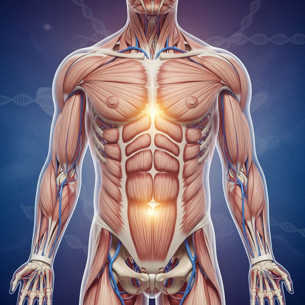
Understanding Human Male Anatomical Development Through Evolution
The human male body exhibits numerous distinctive characteristics that set it apart from other primates, with perhaps none more notable than the unique structure of male genitalia. Unlike many mammalian species that possess a penile bone, humans have evolved a fundamentally different anatomical approach. This evolutionary divergence represents millions of years of natural and sexual selection, with implications for reproduction, mate competition, and physical capability. Understanding the biological reasons behind these anatomical features provides insight into human evolutionary history and the pressures that shaped modern human physiology.
The Absence of Penile Ossification in Humans
Among primates, humans represent an unusual case in lacking a penile bone, or baculum, a structure present in virtually all other mammalian species. This absence is not a disadvantage but rather reflects a specialized evolutionary adaptation. In species possessing an os penis, the bone structure provides mechanical support and rigidity independent of vascular pressure. However, humans have developed an alternative solution that offers distinct advantages for their particular ecological and social context.
The loss of the penile bone coincided with human bipedalism and upright posture. This anatomical change likely provided evolutionary benefits for locomotion, as a rigid bone structure extending from the body would have created a significant impediment to movement and balance in an upright-walking primate. This represents a classic example of evolutionary trade-offs, where the advantages gained in mobility outweighed the mechanical benefits provided by ossification.
To compensate for the absence of bone, humans developed sophisticated tissue architecture. The tunica albuginea, a specialized fibrous sheath surrounding the erectile tissue, contains abundant elastic fibers that maintain rigidity through structural tension rather than skeletal support. Additionally, a specialized ligamentous structure called the distal ligament serves as a supporting framework for the glans, functioning similarly to how a penile bone would in other species. This structural innovation demonstrates how evolution can solve the same functional problems through different anatomical pathways.
The Role of Hemodynamic Erection Mechanisms
Human males achieve erections through a purely hemodynamic process, relying entirely on blood pressure and vascular mechanics rather than mechanical bone structure. This system involves the filling of specialized erectile tissue chambers, the corpora cavernosa, with blood under pressure. The venous architecture plays a critical role in maintaining tumescence by restricting blood outflow through a sophisticated system of veins and fascial layers.
The vascular system supporting erection in humans is remarkably complex. Research has identified multiple venous pathways, including dorsal arteries, para-arterial veins, and cavernosal veins, all precisely arranged within fascial compartments to optimize pressure maintenance. This intricate vascular engineering allows humans to achieve and sustain rigid erections without skeletal support, enabling reproductive function in an upright-posture species.
The hemodynamic system offers advantages beyond basic reproductive capability. It allows for variable penile extensibility, meaning the organ can change length and position without risk of fracture or mechanical failure. This flexibility contributes to the diversity of sexual positioning unique to humans, facilitating the behavioral and social dimensions of human reproduction that extend beyond immediate procreative necessity.
Evolutionary Theories Explaining Genital Morphology
Biologists have proposed several competing theories to explain why human male genitalia evolved their distinctive shape and size relative to other primates. These theories highlight different selective pressures that may have shaped reproductive anatomy over evolutionary time.
The Pleasure and Behavioral Flexibility Hypothesis
One prominent theory suggests that human genital morphology evolved to enhance sexual pleasure and facilitate behavioral diversity. Humans possess larger genitalia relative to body size compared to other primates. Coupled with the absence of penile spines found in many other species, human males must invest significantly more time and effort in sexual activity to achieve climax. This extended duration creates opportunities for pair bonding and increased mate investment, potentially strengthening social partnerships.
The hemodynamic system also permits a remarkable range of sexual positions and activities unavailable to species constrained by skeletal penile structure. This anatomical flexibility may have created selective pressure favoring females who experienced greater pleasure during intercourse, as sexual satisfaction could influence mate choice and pair formation. Over generations, males capable of providing more pleasurable experiences would have enjoyed reproductive advantages, driving the evolution of larger, more responsive genital structures.
The Sperm Competition and Displacement Theory
An alternative evolutionary framework emphasizes reproductive competition between males. The semen displacement theory proposes that human penile anatomy evolved as a specialized tool for sperm competition in contexts of multiple matings. The distinctive mushroom-shaped glans, characterized by a pronounced coronal ridge, may function mechanically during intercourse to displace previously deposited semen from competing males.
This theory emerged from observations that human females, unlike females of many other primate species, do not display obvious fertility signals, and ancestral human mating systems may have involved multiple simultaneous partnerships. Under these conditions, the ability to effectively displace rival sperm would confer substantial reproductive advantages. The internal structure of the glans, with its specialized tissue architecture, may optimize this mechanical function while simultaneously maintaining the structural integrity necessary for repeated sexual activity.
Sexual Selection and Mate Preference
A broader framework incorporating sexual selection recognizes that genital morphology may reflect aesthetic preferences of potential mates. Recent research using life-sized digital models suggests that human perception of male attractiveness is subtly influenced by genital size and shape. Over evolutionary time, if females showed consistent preferences for particular genital configurations, these traits would have become increasingly common in populations through preferential mating.
Sexual selection operates independently from natural selection and can drive traits to exaggerated degrees when mate preference is sufficiently strong. Human genital morphology may represent a balance between competing pressures: the functional requirements for sperm competition, the biomechanical constraints imposed by upright posture, and the aesthetic preferences encoded in female mate choice mechanisms.
Structural Innovations and Tissue Architecture
Beyond broad evolutionary theories, detailed anatomical research has revealed sophisticated structural solutions that enable human reproductive function without penile ossification. These innovations demonstrate how specialized tissues and precise architectural arrangement can substitute for skeletal support.
The Distal Ligament Structure
The distal ligament represents a particularly remarkable evolutionary innovation. This centrally-positioned structure, composed of fibrous tissue with substantial elastic fiber content, acts as a structural trunk for the glans penis. Microscopic analysis reveals that the tissue composition mirrors the histological properties of penile bones found in other mammals, suggesting evolutionary continuity despite the absence of true ossification.
The distal ligament serves multiple critical functions. It maintains patency of the distal urethra, preserving an open passage for ejaculation even during rigid erection. It also protects the reflexogenic erectile mechanism, the neurological systems controlling erectile response, and provides tensile strength capable of withstanding the buckling pressures generated during intercourse. Remarkably, experimental studies demonstrate that even after partial venous removal, penile rigidity can be maintained through the structural support provided by this ligament, evidence of its mechanical robustness.
Fascial Layering and Compartmentalization
Human penile anatomy incorporates multiple fascial layers that create distinct compartments optimizing pressure maintenance and structural stability. The tunica albuginea, a tough fibrous sheath, completely encloses the erectile tissue chambers while remaining anchored to surrounding structures through ligamentous attachments. Buck’s fascia and Colles’ fascia provide additional protective layers, separating the erectile bodies from skin and dermis.
Within the corpora cavernosa, the paired erectile tissue chambers, a sophisticated arrangement of intracavernosal pillars creates internal structural compartments that facilitate rigidity through chamber design. The median septum, which separates the two corpora cavernosa, demonstrates variable completeness with fenestration patterns correlating to the number and distribution of intracavernosal pillars. This variable architecture appears optimized to balance tensile capability with pressure maintenance across different penile regions.
Specialized Anatomy of the Glans
The glans penis, the distinctive enlarged head of the penis, exhibits unique anatomical properties distinct from the penile shaft. The glans features a mono-layered tunica albuginea and lacks the complete outer longitudinal layer found in the shaft, providing structural properties suited to its particular functions. This modified architecture allows the glans to maintain its expanded shape and structural integrity while accommodating the passage of semen through the urethra during ejaculation.
The mushroom-shaped appearance characteristic of human male genitalia results from this differential tissue architecture. The coronal ridge, the prominent circumferential edge where the glans expands beyond the penile shaft, emerges from the architectural differences between glans and shaft tissues. This distinctive morphology appears optimized for both structural integrity during coitus and mechanical effectiveness in sperm displacement scenarios.
Comparative Primate Anatomy and Evolutionary Context
Understanding human genital evolution requires comparison with related primate species. Among primates, humans, woolly monkeys, and spider monkeys uniquely lack a penile bone. This exceptional pattern suggests that specific evolutionary pressures in these lineages created selective advantages favoring hemodynamic erectile systems over ossified structures.
Most other mammals, including the vast majority of primates, retain penile bones that provide mechanical rigidity independent of vascular pressure. Animals with os penis structures can achieve erections more rapidly and maintain them with less metabolic demand. They can also sustain high-frequency copulation without extended recovery periods, as exemplified by lions and other species capable of dozens or hundreds of copulations daily.
The evolutionary loss of the penile bone in certain lineages represents a derived condition, meaning these species evolved secondarily away from the ancestral mammalian state. This transition typically occurs only when alternative anatomical solutions provide sufficient functional advantages to offset the loss of skeletal support. The hemodynamic system in humans and the few other primates lacking os penis appears to satisfy these evolutionary requirements through sophisticated vascular and connective tissue architecture.
Functional Implications and Reproductive Advantage
The distinctive morphology of human male genitalia provides several functional advantages relevant to human reproductive ecology and behavior. These advantages help explain why these anatomical features persisted and became more pronounced through evolutionary time rather than reverting to more typical primate configurations.
- Extended sexual activity duration through reduced penile spine stimulation, facilitating partner bonding and satisfaction
- Mechanical effectiveness in sperm displacement during scenarios of reproductive competition
- Behavioral flexibility enabling diverse sexual positioning and activity patterns
- Compatibility with bipedal locomotion through anatomical adaptation to upright posture
- Variable extensibility permitting accommodation to different body positions without mechanical failure
- Structural capacity to withstand repeated sexual activity and pressure loads without bone fracture
Clinical and Medical Implications
Understanding penile anatomy has important applications in clinical medicine and surgical intervention. Physicians treating erectile dysfunction, penile trauma, or performing reconstructive procedures benefit from detailed knowledge of tissue architecture and functional mechanics. The discovery of the distal ligament’s structural properties, for instance, has informed surgical approaches to penile venous insufficiency and erectile dysfunction treatment.
The complex vascular anatomy, including the multiple venous pathways previously underestimated by classical anatomy, provides better targets for surgical intervention and improved understanding of hemodynamic erectile physiology. Recognition that rigid erection can be maintained even after partial venous removal demonstrates the robustness of penile structural design and informs realistic expectations for surgical outcomes.
Frequently Asked Questions
Why do humans lack a penile bone when most other mammals have one?
Humans likely lost the penile bone through evolutionary pressures favoring upright bipedal locomotion. A rigid bone structure extending from the body would have impeded movement and balance. Instead, humans developed sophisticated hemodynamic and connective tissue systems providing equivalent functional benefits without the mechanical impediment.
How does the hemodynamic erection system work without a bone?
Human erections rely on blood pressure filling specialized erectile tissue chambers (corpora cavernosa) while venous drainage is restricted through fascial compartmentalization. Elastic fibers in the tunica albuginea and the distal ligament maintain structural rigidity. Intracavernosal pillars and median septum fenestration optimize pressure distribution and tensile capability.
What is the distal ligament and why is it important?
The distal ligament is a specialized fibrous structure with elastic fibers that supports the glans penis, maintains urethral patency, and protects the erectile nerve mechanisms. It functions similarly to a penile bone in other species, providing structural support and demonstrating the robustness of human penile architecture through hemodynamic mechanisms.
Does the mushroom shape of the glans serve a specific function?
The distinctive mushroom morphology may serve multiple functions including mechanical effectiveness in sperm displacement, structural optimization for maintaining rigidity during coitus, and potential aesthetic appeal influencing mate selection. The coronal ridge and expanded glans architecture emerge from tissue composition differences suited to these combined functional requirements.
How did scientists discover the complex penile vascular anatomy?
Detailed vascular anatomy was revealed through dissection and histological analysis of human cadavers, along with hemodynamic studies on fresh and preserved specimens. Researchers identified seven distinct veins between the tunica albuginea and Buck’s fascia, far exceeding the single deep dorsal vein previously documented in textbooks.
Conclusion: Evolution as Anatomical Solution
The distinctive anatomy of human male genitalia represents an elegant evolutionary solution to multiple competing pressures. Rather than retaining the penile bone characteristic of ancestral mammals, humans evolved a sophisticated hemodynamic system supported by specialized connective tissues and fascial compartmentalization. This transformation occurred as selective pressures related to upright posture, reproductive competition, and behavioral flexibility shaped human morphology over millions of years.
The mushroom-shaped glans, the hemodynamic erectile mechanism, the distal ligament, and the complex vascular architecture all work together as an integrated system optimized for human reproductive function and locomotion. Evolutionary biologists continue to refine understanding of precisely which selective pressures drove these anatomical changes, with evidence supporting multiple competing theories about pleasure enhancement, sperm competition, and sexual selection. Regardless of which mechanisms dominated, the result demonstrates how evolution can achieve sophisticated functional outcomes through novel anatomical arrangements rather than simply copying solutions from ancestral species.
References
- Advances in understanding of mammalian penile evolution, human penile anatomy and human erection physiology: clinical implications for physicians and surgeons — National Center for Biotechnology Information (NCBI). PMC3560785. https://pmc.ncbi.nlm.nih.gov/articles/PMC3560785/
- How Did Man Lose His Penis Bone? — UCL Researchers in Museums. 2012-11-26. https://blogs.ucl.ac.uk/researchers-in-museums/tag/human-penile-evolution/
- The History Of The Penis — Educational Media. https://www.youtube.com/watch?v=hFF0fOaS2QA
- The long and short of it: Human penis size evolved for both attraction and rivalry — News Medical Life Sciences. 2026-01-25. https://www.news-medical.net/news/20260125/The-long-and-short-of-it-Human-penis-size-evolved-for-both-attraction-and-rivalry-study-suggests.aspx