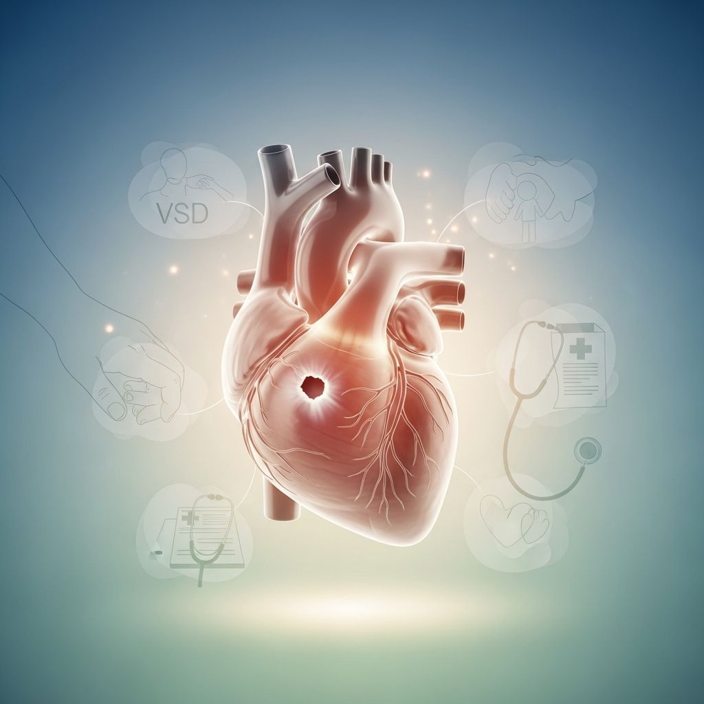
Ventricular septal defect (VSD) is one of the most common heart differences present at birth, and it happens when a baby is born with a small opening between the heart’s lower chambers. Although the word “defect” can sound frightening, many VSDs are small, cause few or no symptoms, and may even close on their own as a child grows. Understanding how VSD works helps parents make confident decisions about tests, treatment, and day-to-day care.
How the Healthy Heart Works
To understand VSD, it helps to first picture a normal heart. The heart is a strong muscle pump with four chambers that move blood in one direction: from the body to the lungs for oxygen, and then back out to the body again.
- The two upper chambers are called atria and receive blood returning from the body and lungs.
- The two lower chambers are called ventricles and pump blood out to the lungs and the rest of the body.
- A wall (septum) separates the right and left sides so that oxygen-poor blood and oxygen-rich blood do not mix.
The right ventricle sends oxygen-poor blood to the lungs, while the left ventricle sends oxygen-rich blood to the body. The septum between the ventricles makes sure blood takes the correct path and prevents unnecessary extra work for the heart.
What Is a Ventricular Septal Defect?
A ventricular septal defect is a hole in the wall that divides the left ventricle from the right ventricle. Because the pressure is higher on the left side, blood usually flows through the opening from the left ventricle into the right ventricle instead of going straight out to the body.
This “extra” flow means the lungs receive more blood than usual, and the heart has to work harder to keep up. The size and location of the opening determine how much extra blood flows and how serious the problem is.
Types of VSD by Size and Location
Doctors often describe VSDs based on where they sit in the ventricular septum and how large they are. These factors guide treatment decisions and long-term follow-up.
By Size
- Small VSD: Allows only a small amount of blood to pass through; often causes a heart murmur but few or no symptoms.
- Medium VSD: Lets more blood flow between the ventricles and may cause faster breathing, poor weight gain, or tiredness in babies.
- Large VSD: Creates a big opening, causing significant extra blood flow to the lungs and putting strain on the heart; symptoms usually appear early in life.
By Location
The ventricular septum is not a flat, uniform wall; it has several regions, and a VSD can occur in different parts of that wall. Each location comes with slightly different surgical approaches and follow-up considerations.
| Feature | Small VSD | Medium / Large VSD |
|---|---|---|
| Typical symptoms | Often none; may only have a heart murmur | Fast breathing, sweating with feeds, poor growth, tiredness |
| Effect on lungs | Little or no extra blood flow | Significant extra blood flow; lungs may become congested |
| Likelihood of closing on its own | Relatively high | Lower; may require surgery or catheter procedure |
| Typical management | Observation and regular checkups | Medication and possible surgical repair |
Why VSD Happens
VSD is a congenital heart difference, meaning it develops while the baby is growing in the womb. Early in pregnancy, the heart starts as a simple tube and gradually forms walls and chambers; if that wall between the ventricles does not fully close, a VSD can remain.
In many families, there is no obvious cause. Sometimes VSD occurs as part of a genetic syndrome or alongside other heart differences, but in most cases parents did nothing to cause the condition and could not have prevented it.
How VSD Affects the Heart and Lungs
When blood flows from the left ventricle to the right ventricle through a VSD, the right side receives extra oxygen-rich blood. That blood is then pumped again to the lungs, which means the lungs receive more blood than they actually need.
- The heart must pump harder, which can lead to enlargement and fatigue of the heart muscle over time.
- The lungs can become overloaded with blood, leading to fast breathing and difficulty feeding.
- If untreated in severe cases, long-term high pressure in the lungs can cause permanent damage to lung blood vessels.
The body’s response depends largely on how big the hole is and how much extra blood is crossing the septum. This is why two children with the same diagnosis of VSD can look very different in daily life.
Signs and Symptoms Parents Might Notice
Some children with a VSD look and act completely healthy, while others show clear signs of heart strain in the first weeks or months of life. The symptoms usually reflect how hard the heart and lungs are working to handle the extra blood flow.
- Fast breathing, especially during feeding or when crying.
- Tiring easily while feeding or playing.
- Sweating on the head or body during feeds, even in a cool room.
- Poor weight gain despite good appetite or frequent feeds.
- Pale or slightly bluish color around the lips or fingernails in more serious cases.
- Frequent respiratory infections, such as colds or chest congestion.
Some babies with small VSDs have no outward symptoms, and the only clue is a heart murmur that a clinician hears with a stethoscope during a routine checkup. For this reason, keeping well-baby and yearly visits is vital even when a child appears healthy.
How VSD Is Diagnosed
The diagnostic process often begins when a healthcare provider hears an unusual sound, called a murmur, while listening to the heart. A murmur is simply the sound of blood flowing in a way that creates extra noise; it can be harmless or related to a structural difference like a VSD.
To understand the cause of the murmur and measure the size and impact of a VSD, doctors may use several tests. These tests are painless or only mildly uncomfortable and are commonly done in children of all ages.
- Echocardiogram (echo): An ultrasound of the heart that shows the structure, the opening in the septum, and how blood flows through the chambers.
- Electrocardiogram (ECG or EKG): Measures the electrical activity of the heart to look for signs of enlargement or strain.
- Chest X-ray: Can show whether the heart looks enlarged or whether there is extra blood flow to the lungs.
- Pulse oximetry: A small sensor on a finger or toe that checks the level of oxygen in the blood.
In more complex situations, additional imaging or a heart catheterization may be needed to measure pressures inside the heart and lungs. The cardiology team uses all of this information to design the safest, most effective care plan.
Treatment Options for VSD
Not every VSD needs active treatment; some only require careful monitoring. The main goals of treatment are to reduce symptoms, protect the lungs and heart from long-term damage, and close or control the hole when necessary.
Watchful Waiting and Regular Checkups
For small VSDs that are not causing symptoms, doctors often recommend observation. Children return for periodic echocardiograms and exams to see whether the VSD is shrinking, closing, or staying the same.
- Parents may be asked to track growth, feeding patterns, and activity levels.
- Yearly or more frequent follow-up allows the doctor to catch any new issues early.
Medications
Medicine does not close the hole itself, but it can ease symptoms while a child grows or waits for a procedure. These medications help the heart and lungs handle the extra workload more comfortably.
- Diuretics: Help the body get rid of extra fluid, making it easier for the lungs to function and reducing swelling.
- Medications that help the heart pump: Can support the heart muscle so it works more efficiently.
- Nutrition support: High-calorie formulas or supplements to help babies gain weight when feeding is tiring.
Surgical Repair
If a VSD is large, causes significant symptoms, or risks damaging the lungs over time, surgical closure may be recommended. The procedure is usually done under general anesthesia by a pediatric heart surgeon in a specialized center.
- The surgeon reaches the heart, opens it, and closes the VSD using a patch or stitches.
- After surgery, most children stay in the hospital for several days for monitoring and recovery.
- Many children experience a clear improvement in breathing, feeding, and energy within weeks.
Catheter-Based Closure
In selected cases, especially for certain types of VSD and in older children, a catheter-based procedure may be an option. A thin tube is threaded through a blood vessel up to the heart, and a device is placed to plug the opening.
Not every child is a candidate for this approach; the decision depends on the VSD’s size, location, and the child’s overall health. The cardiology team will discuss the best method and explain the benefits and risks of each.
Possible Complications and Long-Term Outlook
When managed appropriately, many children with VSD grow up to lead active, healthy lives. Still, it is important to understand potential complications so parents know why follow-up care is essential.
- Heart failure: In severe cases, the heart can become too weak or stiff to pump effectively if the VSD is not treated.
- Pulmonary hypertension: Long-term extra blood flow to the lungs can raise pressure in the lung arteries, potentially causing lasting damage.
- Heart rhythm problems: Some children can develop irregular heartbeats and may need monitoring or medication.
- Heart valve issues: Extra flow can affect nearby valves, sometimes leading to leakage that requires monitoring.
- Infection inside the heart (endocarditis): A small number of children with VSD may have a higher risk of infection on the inner lining of the heart, particularly if they also have abnormal valves or past surgery.
The risk of these problems is much lower when VSDs are found early, monitored regularly, and treated as needed. Many children require lifelong cardiology follow-up, but their day-to-day activities can often be very similar to those of other children.
Daily Life, Activities, and School
Parents often want to know how VSD will affect their child’s ability to play, attend school, and live a typical childhood. The answer depends on the size of the VSD, whether it has been repaired, and how well the heart and lungs are functioning.
- Many children with small or repaired VSDs can participate in regular physical activity without special limits.
- Some children with ongoing heart or lung strain may need temporary restrictions on intense sports until cleared by their cardiologist.
- Teachers and school nurses should be told about the heart condition and any medications or activity guidelines.
Good sleep, balanced nutrition, and routine vaccinations are all important for overall health. Emotional support for both the child and family members is also key, especially around the time of tests, surgery, or hospital stays.
Supporting Your Child Emotionally
Medical visits, tests, and procedures can feel overwhelming for children and parents alike. Building a calm, informed approach can help reduce worry and help children feel more secure.
- Use simple, age-appropriate language to explain what is happening and why.
- Offer choices where possible (which toy to bring, which arm for a blood pressure cuff) to give a sense of control.
- Stay in close communication with the healthcare team and ask questions until the plan feels clear.
- Consider connecting with other families who have children with congenital heart differences for peer support.
Frequently Asked Questions About VSD
Will my child outgrow a VSD?
Some children, especially those with small VSDs, may experience natural closure as the heart grows and the septum thickens. Others will continue to have a small opening that does not cause significant problems and simply needs periodic monitoring.
Is VSD caused by something I did during pregnancy?
In most cases, no specific action or event during pregnancy can be blamed for VSD. The heart forms very early in development, and subtle changes in growth pathways can lead to a small opening even when parents did everything recommended for a healthy pregnancy.
Can my child play sports?
Many children with small or successfully repaired VSDs can safely participate in sports and physical education. A cardiologist will review test results and provide clear guidance on which activities are safe and whether any limits are necessary.
Will my child always need heart medicines?
Medications for VSD are often temporary and used to relieve symptoms, especially before or shortly after surgery. Some children eventually no longer need them, while others with more complex heart differences may require longer-term treatment; this will be discussed at follow-up visits.
What should I do if my child seems worse?
Parents should call their child’s healthcare team right away if they notice increasing breathing trouble, unusual sleepiness, bluish coloring, or sudden changes in feeding or activity. In an emergency, seek urgent medical care or call local emergency services immediately.
Key Takeaways for Parents
- Ventricular septal defect is a common congenital heart difference in which a hole exists between the heart’s lower chambers.
- Many VSDs are small and cause few problems, while larger ones may require medications or surgery to protect the heart and lungs.
- Regular checkups with a pediatric cardiologist, attention to symptoms, and supportive home care give children with VSD an excellent chance to thrive.