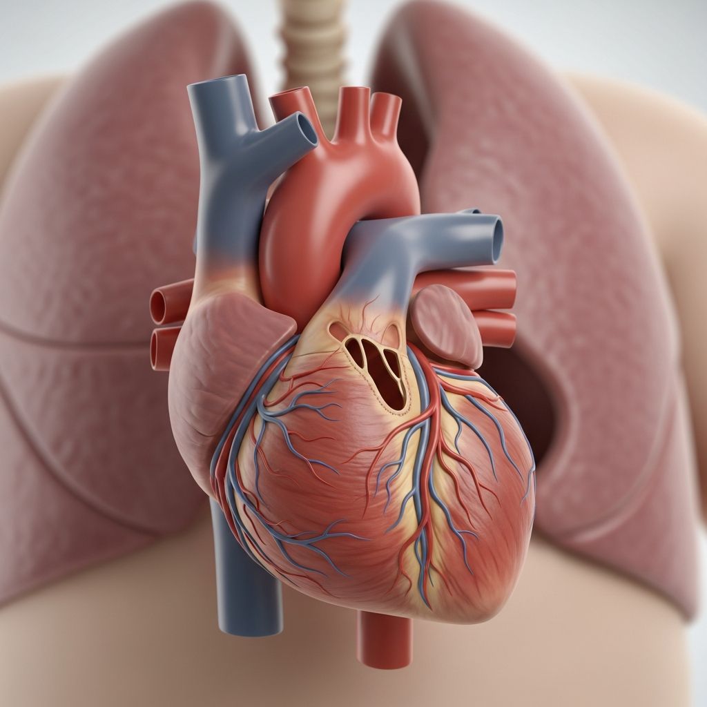
This rare congenital heart condition affects how blood leaves the heart, leading to oxygen shortages and overcirculation to the lungs. Early detection and surgery are vital for survival and development.
The Heart’s Normal Development and What Goes Wrong
In typical fetal growth, the heart forms two distinct major arteries: the aorta for body circulation and the pulmonary artery for lung oxygenation. These emerge from separate ventricular chambers, ensuring oxygen-poor blood goes to the lungs and oxygen-rich blood nourishes the body.
With **truncus arteriosus**, this division fails. A single large trunk, called the truncus arteriosus, arises from both ventricles, overriding a large opening known as a ventricular septal defect (VSD). Oxygenated and deoxygenated blood mix freely, resulting in insufficient oxygen delivery body-wide while excess blood floods the lungs.
This defect arises early in pregnancy when the embryonic truncus—a temporary single vessel—does not properly split. Nearly all cases include a VSD, and the truncal valve may be malformed, allowing backward leakage (regurgitation).
Recognizing Early Warning Signs in Newborns
Symptoms emerge rapidly after birth as the baby transitions from placental to lung-based oxygenation. Common indicators include:
- Bluish skin tint (cyanosis), especially around lips and nails, due to low oxygen levels.
- Rapid, labored breathing or frequent respiratory distress.
- Poor feeding, excessive sweating during meals, and failure to thrive with slow weight gain.
- Heart murmur audible via stethoscope from turbulent blood flow.
- Enlarged liver from heart strain backing up circulation.
Without prompt care, these progress to full heart failure, marked by extreme fatigue and fluid buildup.
Associated Conditions and Genetic Links
Truncus arteriosus rarely stands alone. About one-third of cases link to DiGeorge syndrome (22q11.2 deletion), involving immune issues, palate anomalies, and learning challenges.
Other frequent companions:
| Associated Defect | Impact |
|---|---|
| Ventricular Septal Defect (VSD) | Enables blood mixing; present in 99% of cases. |
| Truncal Valve Abnormalities | Regurgitation strains the heart. |
| Interrupted Aortic Arch | Blocks body blood flow; requires urgent meds. |
| Pulmonary Artery Variations | Alters lung blood supply. |
Family history of heart defects or syndromes raises risk, though most cases are sporadic.
How Specialists Confirm the Diagnosis
Prenatal ultrasound may hint at issues, but postnatal tests confirm. Key diagnostics:
- Echocardiogram: Gold standard; visualizes the single trunk, VSD, and valve function via ultrasound waves.
- Chest X-ray: Reveals enlarged heart and excess lung fluid.
- ECG: Detects enlarged chambers and irregular rhythms.
- Cardiac Catheterization: Measures pressures and oxygen levels; guides surgery.
Genetic testing screens for DiGeorge and others, aiding holistic care.
Life-Saving Surgical Interventions
Surgery within the first weeks is essential, as untreated mortality exceeds 80% in infancy.
Core Procedure: Surgeons close the VSD, detach pulmonary arteries from the truncus (fashioning a new right ventricle-to-pulmonary conduit), and reconstruct the aorta with its valve. A valved homograft or conduit ensures proper pulmonary flow.
Variations depend on anatomy; interrupted arch needs additional aorta repair.
Post-op, infants stabilize in ICU with ventilators and meds. Hospital stays span weeks, with feeding support for growth.
Navigating Recovery and Long-Term Care
Initial recovery focuses on heart function stabilization. Medications like diuretics reduce fluid, while ACE inhibitors ease pumping.
Long-term realities include:
- Conduit and valve replacements every 10-15 years due to growth/outwear.
- Regular cardiology visits monitoring leaks, blockages, arrhythmias.
- Activity limits initially; most lead active lives post-repair.
- Endocarditis prevention with antibiotics before dental work.
Neurodevelopmental support addresses potential delays from oxygenation issues or syndromes.
Outlook and Factors Influencing Prognosis
Survival has soared to 80-90% at 20 years with timely surgery at expert centers. Key influencers:
- Surgery timing: Earlier is better.
- Valve condition: Severe initial regurgitation worsens outlook.
- Genetics: DiGeorge adds complexities.
- Follow-up adherence: Prevents complications.
Adults often need reoperations but enjoy good quality of life.
Support Strategies for Families
Caring for a child with truncus arteriosus demands emotional and practical resilience. Join support groups via ACHA or local chapters. Track milestones, advocate in schools, and prioritize mental health.
Genetic counseling clarifies recurrence risks (1-3% for siblings).
Frequently Asked Questions (FAQs)
Can truncus arteriosus be detected before birth?
Yes, fetal echocardiography around 18-22 weeks often identifies the single vessel and VSD, enabling birth planning at specialized centers.
What is the survival rate after surgery?
Over 90% survive the initial operation; 20-year survival nears 85% with ongoing care.
Will my child need multiple heart surgeries?
Typically yes, for conduit/Valve replacements as they grow, usually 2-4 times lifetime.
Are there exercise restrictions?
Contact sports avoided; low-moderate activities encouraged post-clearance.
Is truncus arteriosus hereditary?
Rarely; mostly random, but syndromes like DiGeorge confer higher familial risk.
References
- Truncus arteriosus – Symptoms and causes — Mayo Clinic. 2023-10-12. https://www.mayoclinic.org/diseases-conditions/truncus-arteriosus/symptoms-causes/syc-20364247
- Truncus Arteriosus — StatPearls, NCBI Bookshelf. 2023-07-17. https://www.ncbi.nlm.nih.gov/books/NBK534774/
- Truncus Arteriosus | Congenital Heart Defects (CHDs) — Centers for Disease Control and Prevention (CDC). 2024-05-07. https://www.cdc.gov/heart-defects/about/truncus-arteriosus.html
- Truncus Arteriosus: Symptoms & Treatment — Cleveland Clinic. 2023-11-02. https://my.clevelandclinic.org/health/diseases/14785-truncus-arteriosus
- Truncus Arteriosus — American Heart Association. 2023-08-15. https://www.heart.org/en/health-topics/congenital-heart-defects/about-congenital-heart-defects/truncus-arteriosus
- Truncus Arteriosus — Adult Congenital Heart Association (ACHA). 2022-11-01. https://www.achaheart.org/your-heart/educational-qas/types-of-heart-defects/truncus-arteriosus/
- Truncus Arteriosus — Children’s Hospital of Philadelphia (CHOP). 2024-01-20. https://www.chop.edu/conditions-diseases/truncus-arteriosus