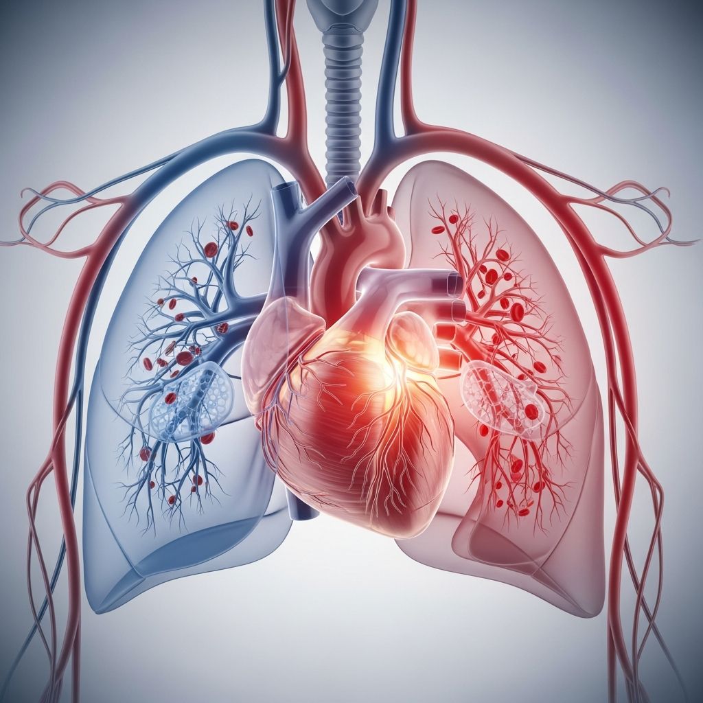
The right ventricle is one of the four chambers of the heart and is responsible for pumping blood from the heart to the lungs so it can pick up oxygen. It sits in the lower right side of the heart and works together with the other chambers and valves to keep blood moving in the correct direction during every heartbeat.
Understanding how the right ventricle works helps parents make sense of heart tests, medical terms, and conditions that might affect a child’s circulation and breathing.
Where the Right Ventricle Sits in the Heart
The heart has two upper chambers (atria) and two lower chambers (ventricles). The right ventricle is the lower chamber on the right side and lies just beneath the right atrium. When viewing the heart from the front, much of what is seen is actually the right ventricle because it forms a large part of the front surface of the heart.
- The right atrium is above it and sends blood down through a valve.
- The left ventricle is next to it and has thicker muscular walls.
- The diaphragm (the main breathing muscle) sits just below the bottom of the right ventricle.
This position allows the right ventricle to act as the main gateway between the heart and the lungs.
What the Right Ventricle Does
The right ventricle’s main job is to move low-oxygen (deoxygenated) blood from the right side of the heart into the lungs. There, the blood receives fresh oxygen and releases carbon dioxide before coming back to the left side of the heart to be pumped out to the body.
Think of the right ventricle as a delivery pump that sends “used” blood to a cleaning and refueling station (the lungs). Without it, blood could not reach the lungs efficiently, and the body’s tissues would quickly run low on oxygen.
The Path of Blood Through the Right Ventricle
During every heartbeat, blood follows a clear path through the right side of the heart before passing to the lungs. The steps below describe this process in simple terms.
- Low-oxygen blood from the body returns to the heart and enters the right atrium.
- When the right atrium squeezes, blood flows down through the tricuspid valve into the right ventricle.
- The right ventricle fills and then contracts, squeezing the blood upward.
- Blood passes through the pulmonary valve and into the pulmonary artery.
- The pulmonary artery carries blood to the lungs, where it picks up oxygen.
Once the blood is oxygen-rich, it returns to the left side of the heart and is then pumped out to the entire body.
Key Structures Inside the Right Ventricle
Although the right ventricle is a hollow chamber, it contains several important structures that support its pumping action and help control blood flow.
- Muscular wall (myocardium): The muscle layer contracts to push blood toward the lungs. It is thinner than the muscle wall of the left ventricle because it does not need to generate as much pressure.
- Tricuspid valve attachments: Small string-like fibers called chordae tendineae connect the tricuspid valve flaps to the inner wall of the right ventricle.
- Papillary muscles: These small muscles anchor the chordae tendineae. When they contract, they help keep the tricuspid valve flaps from swinging backward into the right atrium.
- Outflow tract: The upper part of the right ventricle that leads to the pulmonary valve and pulmonary artery, guiding blood out toward the lungs.
All of these parts must work together smoothly to keep blood moving forward and prevent it from leaking backward.
Right Ventricle vs. Left Ventricle
The right and left ventricles share the same basic role of pumping blood, but they send blood to different places and work under different pressures. The right ventricle sends blood to the lungs, while the left ventricle sends blood to the rest of the body.
This difference in workload explains why the left ventricle has thicker walls and why problems that increase pressure in the lungs can strain the right ventricle.
How the Two Ventricles Differ
| Feature | Right Ventricle | Left Ventricle |
|---|---|---|
| Main destination of blood | Sends blood to the lungs through the pulmonary artery | Sends blood to the body through the aorta |
| Wall thickness | Thinner, because it pumps against low pressure in the lungs | Thicker, because it pumps against higher pressure in the body |
| Type of blood | Handles low-oxygen blood returning from the body | Handles oxygen-rich blood returning from the lungs |
| Main valve at exit | Pulmonary valve | Aortic valve |
Even though the left ventricle has more muscle, both ventricles pump about the same amount of blood each minute to keep the circulation balanced.
Why the Right Ventricle Uses Lower Pressure
The lungs are located close to the heart, and the blood vessels inside them are delicate and thin. The right ventricle only needs to generate enough pressure to move blood through the lungs, not across the whole body. Because of this, it works at a lower pressure than the left ventricle.
If pressure in the lungs rises too high, the right ventricle must work harder, which can cause the muscle to thicken or weaken over time. This is one reason doctors take conditions that raise lung pressure, such as certain lung diseases or heart defects, very seriously.
How the Right Ventricle Works During a Heartbeat
Each heartbeat includes a filling phase and a pumping phase. During the filling phase, the right ventricle relaxes and receives blood from the right atrium. During the pumping phase, it contracts and sends blood toward the lungs.
- Relaxation (diastole): The tricuspid valve is open, the pulmonary valve is closed, and blood flows down from the right atrium.
- Contraction (systole): The tricuspid valve closes to stop backward flow, the pulmonary valve opens, and blood is forced into the pulmonary artery.
This cycle repeats with every beat, usually many times each minute in infants and fewer times per minute in older children and adults.
Conditions That Can Affect the Right Ventricle
The right ventricle can be affected by heart defects present at birth, diseases that change the structure of the heart muscle, or problems that increase pressure in the lung circulation. Some issues involve only the right side, while others affect the heart as a whole.
- Right-sided heart failure: When the right ventricle cannot pump blood effectively, blood may back up into the veins, causing swelling of the legs, abdomen, or around the eyes.
- Pulmonary hypertension: High blood pressure in the lung arteries makes the right ventricle pump against extra resistance, which can lead to enlargement or weakness of the muscle.
- Valve problems: Narrowing or leaking of the pulmonary or tricuspid valve can force the right ventricle to work harder.
- Certain genetic or structural conditions: Some rare disorders change the way the right ventricle muscle is built or how the large vessels connect to it, which may require specialized care and sometimes surgery.
Symptoms may include tiredness, shortness of breath, poor feeding or slow weight gain in babies, swelling, or a bluish color to the lips or fingers. Any of these signs should prompt a conversation with a healthcare professional.
How Doctors Examine the Right Ventricle
Doctors and pediatric cardiologists use different tests to see how well the right ventricle is shaped and how strongly it contracts. These tests help them decide whether treatment is needed and which options are safest.
- Physical exam: Listening with a stethoscope, checking for extra heart sounds, and looking for signs of swelling or color changes.
- Echocardiogram (heart ultrasound): A painless test that uses sound waves to create moving pictures of the heart chambers and valves.
- Electrocardiogram (ECG or EKG): Measures the electrical signals that control the heartbeat and can show patterns that suggest strain or enlargement.
- Additional imaging: In some cases, cardiac MRI or CT scans give more detailed pictures of the right ventricle’s size, shape, and function.
These tools help doctors monitor children with known heart problems and check for changes over time.
Supporting a Healthy Right Ventricle
For most children with a normal heart, the right ventricle functions well without any special attention. However, healthy daily habits still matter because they support the entire cardiovascular system.
- Encouraging age-appropriate physical activity, as advised by a healthcare provider.
- Offering a balanced diet that includes fruits, vegetables, whole grains, and lean protein.
- Keeping up with regular checkups so that blood pressure, growth, and heart sounds can be monitored.
- Following specialist instructions carefully if a child has an existing heart condition.
Parents who understand the basics of heart structure can participate more confidently in decisions about tests, treatments, and lifestyle recommendations.
Simple Word Picture of Blood Flow
It can help to imagine heart and lung circulation as two loops that meet in the right and left ventricles. The right ventricle sits between the body and the lungs, while the left ventricle sits between the lungs and the rest of the body.
- Loop 1: Body → right atrium → right ventricle → lungs.
- Loop 2: Lungs → left atrium → left ventricle → body.
The right ventricle is the pump that pushes blood through the first loop, making sure the lungs always receive the blood they need to refresh.
Frequently Asked Questions About the Right Ventricle
Is the right ventricle less important than the left ventricle?
No. While the left ventricle has a thicker muscle wall and sends blood to the whole body, the right ventricle is just as essential. Without it, blood would not reach the lungs to receive oxygen, and the body would not function for long.
Can children have problems specifically with the right ventricle?
Yes. Some heart defects present at birth and some rare muscle conditions mainly affect the right ventricle or the valves that connect to it. These conditions are often managed by pediatric cardiologists using a mix of medicines, procedures, and sometimes surgery.
What symptoms might suggest a right-sided heart issue?
Warning signs can include getting tired easily, trouble breathing, swelling of the legs or belly, poor feeding in babies, or a bluish color of the lips or fingernails. Any of these signs should be discussed promptly with a healthcare provider.
How can parents support a child with a right ventricle condition?
Following the care plan, keeping all specialist appointments, giving medications as directed, and reporting any new or worsening symptoms are the most important steps. Emotional support, clear routines, and age-appropriate activity also help children cope better with ongoing heart care.
Does exercise harm the right ventricle?
For children with a normal heart, regular movement is healthy and supports both heart and lung function. If a child has a diagnosed heart condition, activity levels should be guided by the cardiologist, who will explain what types and amounts of exercise are safest.
Key Takeaways for Parents
- The right ventricle is the lower right heart chamber that sends blood to the lungs.
- It works at lower pressure than the left ventricle but is equally vital for healthy circulation.
- Problems that raise pressure in the lungs or affect the right-side valves can strain this chamber.
- Understanding the right ventricle’s role makes it easier to follow medical advice and ask informed questions about a child’s heart health.
With a basic grasp of how the right ventricle functions, parents can better understand test results, medical explanations, and treatment plans related to their child’s heart and lung circulation.