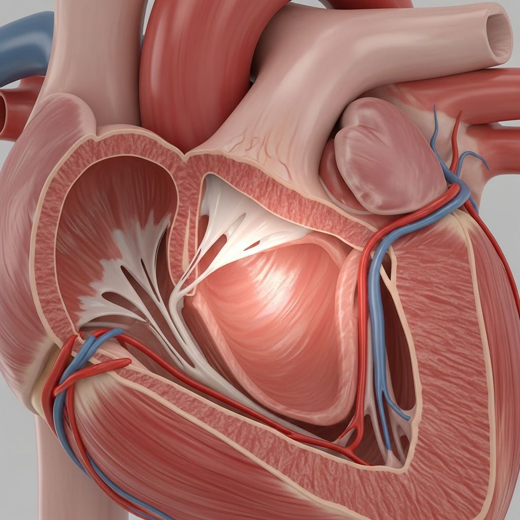
The right atrium is one of the heart’s four chambers and acts as the main entry point for blood returning from the body. It may not receive as much attention as the heart’s pumping chambers, but without the right atrium, normal blood circulation would not be possible.
Understanding how the right atrium works helps make sense of blood pressure, heart rhythm, and many common heart conditions. This guide explains its structure, function, and everyday importance in clear, family-friendly language.
Where the Right Atrium Sits in the Heart
The heart is divided into a right side and a left side, each with an upper chamber (atrium) and a lower chamber (ventricle). The right atrium is the upper chamber on the right side of the heart, facing the front of the chest more than the left atrium.
From the outside, the right atrium forms much of the upper right portion of the heart that lies just behind the breastbone. It connects above to the large body veins and below to the right ventricle.
- The right atrium sits above the right ventricle.
- It lies mostly toward the right side of the chest.
- It is attached to the body’s two largest veins and a special vein from the heart muscle itself.
Basic Structure of the Right Atrium
The right atrium is a hollow, muscular chamber with thin walls compared with the ventricles. Its size and shape allow it to serve as a flexible “waiting room” for blood before it moves to the right ventricle.
Although it may look like a simple pocket, the right atrium has several distinct regions and openings that guide blood flow efficiently.
Key Internal Areas
Inside the right atrium, parts of the wall are smooth while other areas contain ridged muscle bands. These design features help the chamber stretch, contract, and direct blood toward the next chamber without turbulence.
- Smooth inflow region: The part of the atrial wall near the openings of the large veins is relatively smooth, allowing blood to enter with minimal resistance.
- Muscular ridges: Bands of muscle fibers along the inner wall help the chamber contract and squeeze blood toward the right ventricle.
- Thin dividing wall: A partition separates the right atrium from the left atrium, preventing mixing of blood between the two sides.
Openings and Connections
Several important blood vessels and valves connect directly to the right atrium. Each has a specific direction of flow so that blood enters and leaves the chamber in an orderly cycle.
| Structure | Role | Direction of Blood Flow |
|---|---|---|
| Superior vena cava | Brings blood from the head, neck, arms, and upper chest | Into the right atrium |
| Inferior vena cava | Returns blood from the abdomen, pelvis, and legs | Into the right atrium |
| Coronary sinus | Carries blood drained from the heart’s own muscle tissue | Into the right atrium |
| Tricuspid valve | Controls blood passing to the right ventricle | Out of the right atrium |
What the Right Atrium Does
The right atrium’s main job is to receive blood that has already traveled through the body and send it on to the right ventricle. This blood is considered “oxygen-poor” because the tissues have used up much of its oxygen supply.
In every heartbeat, the right atrium coordinates with the other chambers so blood can move smoothly in a one-way loop through the heart, lungs, and body.
Step-by-Step: Blood Flow Through the Right Atrium
- Blood returns from the body through the superior and inferior vena cava.
- This blood enters the right atrium and briefly collects there.
- The right atrium gently contracts, pushing blood through the tricuspid valve.
- Blood flows into the right ventricle, which will send it on to the lungs for oxygen.
During this process, valves between chambers act like one-way doors, ensuring that blood moves forward and does not leak backward when the heart squeezes.
The Right Atrium and the Heart’s Electrical System
Beyond acting as a blood reservoir, the right atrium plays a leading role in controlling the heartbeat. Special clusters of cells in its wall generate and relay electrical signals that tell the heart when to contract.
These cells work like a built-in pacemaker and wiring system, helping maintain a steady rhythm or adjusting the heart rate when someone exercises, feels stress, or rests.
The Natural Pacemaker
Near the opening of the large upper vein sits the heart’s primary pacemaker, a small area of specialized muscle cells. These cells automatically fire electrical impulses without any conscious effort.
- They set the basic heart rate at rest.
- They speed up when the body needs more oxygen, such as during exercise.
- They slow down during sleep or relaxation。
The electrical signal created in this region spreads across both atria, causing them to contract and push blood into the ventricles. The signal then travels to another relay point near the lower part of the right atrium before continuing into the ventricles.
Right Atrium in the Circulatory Loop
The right atrium helps link the systemic circulation (blood going to and from the body) with the pulmonary circulation (blood going to and from the lungs). Its position at this crossroads keeps the two sides of the circulation coordinated.
By adjusting how much blood moves into the right ventricle with each beat, the right atrium helps match the flow coming from the body with the flow going out to the lungs.
- Systemic veins return oxygen-poor blood to the right atrium.
- The right ventricle sends this blood to the lungs for oxygen pickup.
- Oxygen-rich blood later returns to the left side of the heart to be pumped back to the body.
How the Right Atrium Changes from Fetus to Newborn
Before birth, a baby’s lungs are not yet used for breathing, so blood follows a different pattern. In the fetal heart, there is a small opening in the wall between the right and left atria that allows blood to bypass the lungs.
After birth, when the baby begins to breathe air, this opening normally closes, and the right atrium takes on its lifelong role as the main gate for blood returning from the body.
Fetal vs. Adult Right Atrium
| Feature | Before Birth | After Birth |
|---|---|---|
| Atrial wall opening | Present to allow blood to pass from right atrium to left atrium | Closes in most people, leaving a small scar or depression |
| Blood use of lungs | Lungs are not used for oxygen exchange | Lungs become the main source of oxygen for the blood |
| Main purpose of right atrium | Part of a special detour route around the lungs | Primary receiving chamber for blood returning from the body |
Common Problems That Affect the Right Atrium
Several heart conditions can involve the right atrium directly or indirectly. Some change its size or shape, while others disrupt its electrical activity or the flow of blood through its valves.
Right Atrial Enlargement
When the right side of the heart has to push against higher than normal pressure in the lungs or the tricuspid valve is narrowed, the right atrium may stretch and enlarge over time. This enlargement can show up on heart imaging tests or electrocardiograms.
- Long-term lung disease can raise pressure in the lung arteries, straining the right side of the heart.
- Valve problems can make it harder for blood to leave the right atrium.
- Some heart birth defects can increase blood flow into the right atrium.
Rhythm Disturbances
Because the heart’s natural pacemaker and key electrical pathways lie within or near the right atrium, changes in this chamber can trigger irregular rhythms. Some rhythms start with extra beats, while others cause the atria to beat very fast and out of sync with the ventricles.
These rhythm problems may cause sensations of fluttering, pounding, or skipped beats. In some cases, people may feel dizzy, short of breath, or unusually tired.
Issues with the Tricuspid Valve
The valve between the right atrium and right ventricle must open and close at the proper times to keep blood moving in one direction. If this valve becomes stiff, leaky, or malformed, the right atrium may have to work harder.
- Narrowing of the valve can prevent blood from leaving the right atrium easily.
- Leakage can send blood backward into the atrium when the ventricle contracts.
- Both situations can lead to symptoms such as swelling in the legs, fatigue, or abdominal discomfort。
Keeping the Right Atrium and Heart Healthy
While many right atrial problems arise from conditions in the lungs, heart valves, or blood vessels, everyday habits still play an important role in protecting this chamber. Choices that support overall heart health also benefit the right atrium.
- Maintain a healthy blood pressure to reduce strain on the heart and blood vessels。
- Avoid tobacco and limit exposure to secondhand smoke to protect the lungs and right side of the heart。
- Stay physically active with age-appropriate movement most days of the week。
- Eat a balanced diet rich in fruits, vegetables, whole grains, and lean proteins。
- Work with a healthcare professional to manage conditions such as sleep apnea, lung disease, or heart valve problems。
When to Ask a Doctor About the Right Atrium
Many people never hear the term “right atrium” until a test result mentions it. Any time a report describes enlargement, thinning, or abnormal rhythm involving this chamber, it is reasonable to ask a clinician to explain what it means in plain language。
Questions about test findings, new symptoms, or treatment options are especially important if there is a history of lung disease, sleep apnea, or heart problems in the family。
SEO-Friendly Summary Bullet Points
- The right atrium is the upper right chamber of the heart and the main entry point for blood returning from the body。
- It receives oxygen-poor blood and passes it through the tricuspid valve to the right ventricle。
- Special cells in the right atrium act as the heart’s natural pacemaker and help regulate heartbeat。
- In fetal life, a small opening between the atria allows blood to bypass the lungs; this usually closes after birth。
- Right atrial enlargement, valve problems, and rhythm disturbances can affect how this chamber works。
Frequently Asked Questions About the Right Atrium
Is the right atrium the same as the right ventricle?
No. The right atrium is the upper chamber that receives returning blood from the body, while the right ventricle is the lower chamber that pumps that blood out to the lungs. They work as a team but have different shapes, wall thicknesses, and jobs。
Why is the right atrium wall thinner than the ventricle wall?
The right atrium only needs to move blood a short distance into the right ventricle at relatively low pressure. Because the right ventricle must push blood all the way through the lungs, it needs a thicker, stronger muscular wall。
What symptoms might point to a problem with the right atrium?
Symptoms can vary with the cause but may include heart palpitations, shortness of breath, swelling in the legs or abdomen, fatigue, or chest discomfort. Any new or concerning symptoms should be evaluated promptly by a healthcare professional。
Can lifestyle changes improve right atrial health?
Healthy daily habits such as not smoking, staying active, managing weight, and following medical advice for blood pressure, lung conditions, and sleep apnea can ease the workload on the right atrium and support better overall heart function。
How are right atrial problems diagnosed?
Clinicians use tools such as physical exams, electrocardiograms, echocardiograms (heart ultrasound), and sometimes advanced imaging to look at the size, shape, and rhythm of the right atrium. The specific tests chosen depend on a person’s symptoms and medical history。