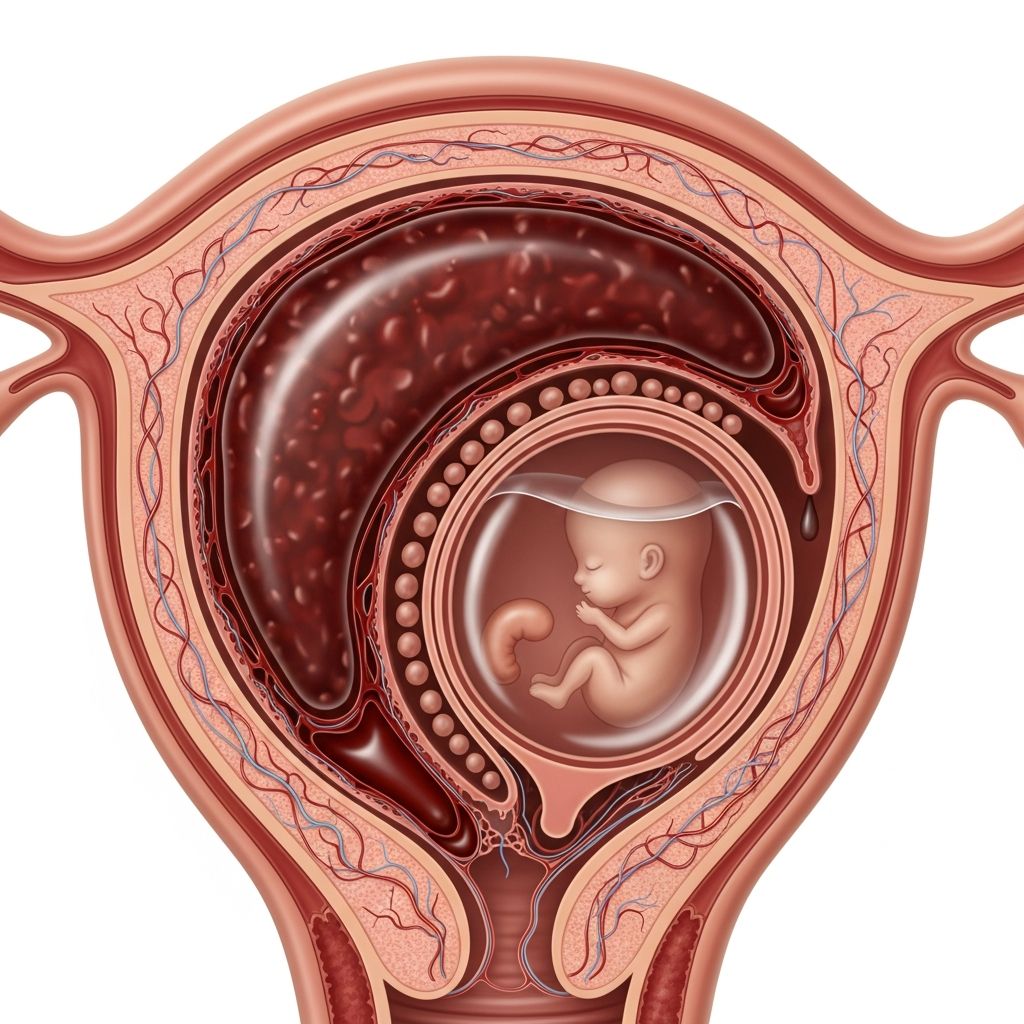
Prenatal ultrasounds use high-frequency sound waves to create images of the developing fetus, offering parents invaluable insights into their baby’s growth and well-being throughout pregnancy. These non-invasive scans help healthcare providers assess key aspects of fetal development while ensuring maternal health.
Why Prenatal Ultrasounds Matter in Modern Pregnancy Care
Ultrasounds have transformed obstetric care by providing real-time visuals of the fetus without radiation exposure. They enable early detection of potential issues, accurate pregnancy dating, and reassurance for parents. Major health organizations like the American College of Obstetricians and Gynecologists (ACOG) endorse their use when medically indicated, balancing benefits against minimal risks.
The primary advantages include confirming viability, identifying multiples, and spotting anomalies early. For instance, scans can reveal placental position, amniotic fluid levels, and fetal heartbeat, guiding decisions on care plans.
When and How Often Are Ultrasounds Performed?
Pregnancies typically involve 2-3 standard ultrasounds, though high-risk cases may require more. Here’s a breakdown:
- First Trimester (8-13 weeks): Confirms heartbeat, measures crown-rump length for dating, screens for chromosomal risks.
- Second Trimester (18-22 weeks): Anatomy scan evaluates organs, limbs, and spine; checks gender if desired.
- Third Trimester (32-36 weeks): Assesses position, growth, and placenta; monitors for issues like growth restriction.
Additional scans may occur for concerns like bleeding, hypertension, or diabetes.
Understanding the Ultrasound Procedure Step-by-Step
Preparation is simple: arrive with a full bladder for early scans to improve image clarity. The sonographer applies gel to your abdomen and glides a transducer over it, capturing images on a screen. The exam lasts 20-45 minutes, is painless, and allows parents to see their baby in motion.
For better views later in pregnancy, a transvaginal probe may be used early on, or you might undress partially. Real-time imaging shows the fetus moving, heart beating, and even yawning, creating memorable moments.
Key Information Revealed by Each Scan
| Scan Timing | Main Purposes | What Parents See/Learn |
|---|---|---|
| Early (6-10 weeks) | Viability, dating, multiples | Gestational sac, yolk sac, fetal pole, heartbeat |
| Mid (18-20 weeks) | Detailed anatomy | Brain, heart, kidneys, spine, limbs; gender option |
| Late (after 28 weeks) | Growth, position | Size estimates, breathing practice, head-down position |
These details help predict due dates accurately (within 5-7 days) and flag issues like ectopic pregnancy or structural defects.
Safety Profile: Are Ultrasounds Risk-Free?
Decades of research affirm ultrasound safety, with no confirmed links to birth defects, cancer, or developmental delays in humans. Mechanisms like thermal heating or cavitation are theoretical but minimized by modern devices adhering to ALARA (As Low As Reasonably Achievable) principles.
Animal studies hint at effects from prolonged exposure, but human data from millions of scans show no increased risks for low birth weight, preterm birth, or childhood issues like dyslexia. A WHO review found no adverse perinatal or long-term outcomes.
Potential Concerns and How They’re Addressed
- Thermal Effects: Heat from waves is limited; operators monitor Thermal Index (TI) below 1.0.
- Mechanical Effects: Micro-bubbles rarely form; Mechanical Index (MI) kept low.
- Doppler Use: Higher energy in first trimester; limit to <60 minutes if needed.
- Non-Medical Scans: Avoid ‘keepsake’ images to prevent extra exposure.
Prudent use by trained professionals ensures benefits outweigh negligible risks.
Interpreting Results: Normal vs. Abnormal Findings
Most scans (95%) are normal, showing expected growth. Abnormalities like shortened limbs or heart defects prompt further tests (e.g., amniocentesis). Measurements compare to percentiles; e.g., head circumference above 90th may indicate macrosomia.
Parents receive preliminary findings immediately, with full reports from physicians. Soft markers (e.g., bright bowel) often resolve harmlessly.
Special Circumstances Calling for Extra Scans
High-risk pregnancies—IVF, maternal age >35, obesity, or conditions like preeclampsia—warrant frequent monitoring. Twins require biweekly checks for twin-to-twin transfusion risks.
Biophysical profiles combine ultrasound with non-stress tests to score fetal well-being on five criteria: movement, tone, breathing, fluid, and heart rate.
Preparing Yourself and Your Partner for the Experience
Discuss expectations with your provider. Bring questions; record the screen if allowed. Emotional responses vary—joy from seeing kicks or anxiety from unknowns. Partner involvement strengthens bonding.
Wear comfortable clothes; fast if instructed for late scans. Post-scan, hydrate and note any provided images.
Advances in Ultrasound Technology Enhancing Care
3D/4D imaging offers detailed, lifelike views for anomaly detection. Doppler assesses blood flow in umbilical cord or heart. AI now aids automated measurements, improving accuracy.
Portable devices enable bedside scans, vital in remote areas.
Frequently Asked Questions (FAQs)
Is it safe to have multiple ultrasounds during pregnancy?
Yes, when medically necessary. Studies confirm no harm from routine scans.
Can ultrasounds determine my baby’s gender accurately?
Typically from 18 weeks, with 95%+ accuracy; external genitals are visible.
What if the ultrasound shows something concerning?
Your doctor will explain and recommend next steps like specialist consults.
Should I avoid ultrasounds for fear of radiation?
No radiation is used—only sound waves, making it safer than X-rays.
How accurate are due dates from ultrasound?
Very precise in first trimester (±5 days); less so later.
Are 3D ultrasounds better than 2D?
They provide clearer images for specific diagnostics, not routine use.
Empowering Parents: Making Informed Decisions
Prenatal ultrasounds bridge the gap between uncertainty and knowledge, fostering proactive care. Discuss options with providers to align with your values, ensuring a healthy journey to parenthood.
References
- Effects of Ultrasound on Biological Tissues and Cells — Global Library of Women’s Medicine (GLOWM). 2012. https://www.glowm.com/article/heading/vol-5–surveillance-of-fetal-wellbeing–effects-of-ultrasound-on-biological-tissues-and-cells/id/411403
- Benefits and risks of ultrasound in pregnancy — PubMed (NCBI). 2013-11-15. https://pubmed.ncbi.nlm.nih.gov/24176149/
- Prudent Use and Safety of Diagnostic Ultrasound in Pregnancy — American Institute of Ultrasound in Medicine (AIUM). 2020-05-19. https://www.aium.org/resources/official-statements/view/prudent-use-and-safety-of-diagnostic-ultrasound-in-pregnancy
- Pregnancy without ultrasound? Pros and cons — UT Southwestern Medical Center. N/A. https://utswmed.org/medblog/pregnancy-without-ultrasound-pros-and-cons/
- Ultrasound In Pregnancy: What To Expect, Purpose & Results — Cleveland Clinic. 2023. https://my.clevelandclinic.org/health/diagnostics/9704-ultrasound-in-pregnancy
- Ultrasound Imaging — U.S. Food and Drug Administration (FDA). 2023. https://www.fda.gov/radiation-emitting-products/medical-imaging/ultrasound-imaging
- Ultrasound — Mayo Clinic. 2023-08-25. https://www.mayoclinic.org/tests-procedures/ultrasound/about/pac-20395177
- Obstetric Ultrasound — RadiologyInfo.org (RSNA). 2023. https://www.radiologyinfo.org/en/info/obstetricus