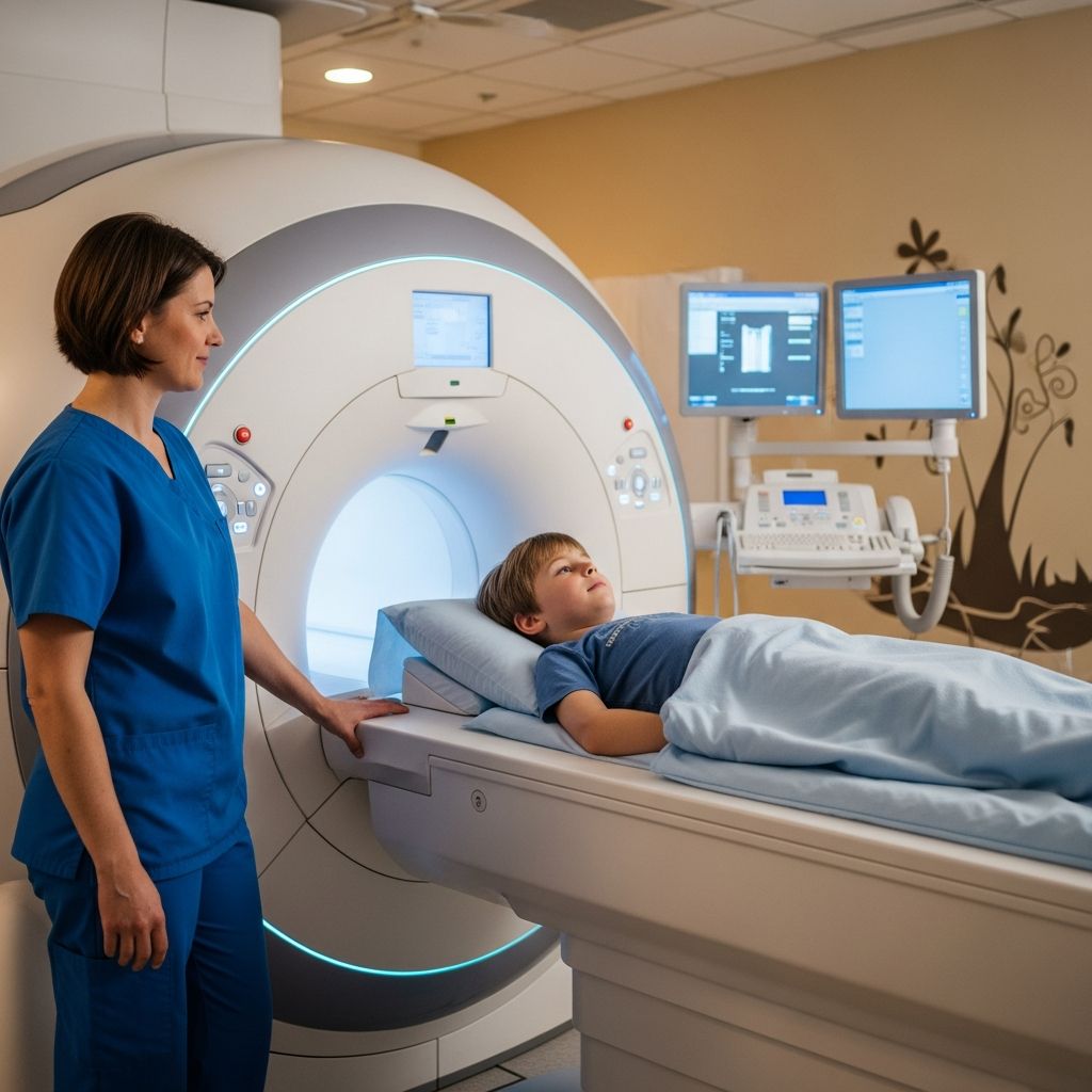
Spinal magnetic resonance imaging (MRI) is a non-invasive diagnostic tool that provides detailed images of a child’s spine, helping doctors identify issues without using radiation. This guide explains the process, reasons for the test, and tips for parents to support their child.
Understanding Spinal MRI in Pediatric Care
Magnetic resonance imaging uses powerful magnets and radio waves to create high-resolution pictures of the spine’s bones, discs, nerves, and surrounding tissues. In children, spinal MRIs are tailored to unique conditions like congenital anomalies or growth-related problems, differing from adult protocols which often focus on degenerative issues.
Pediatric spine imaging addresses a broad range of indications, including scoliosis, cord tethering, Chiari malformations, tumors, trauma, and non-accidental injuries. Protocols are customized to the clinical question to ensure accurate diagnosis and avoid unnecessary scans.
Common Reasons for Recommending a Spinal MRI
Doctors order spinal MRIs for children when symptoms suggest underlying spinal problems that X-rays or other tests can’t fully reveal. Key indications include:
- Persistent back or leg pain: Especially if accompanied by numbness, weakness, or bowel/bladder issues, indicating possible disc herniation or spinal stenosis.
- Abnormal spine curvature: Such as scoliosis, to check for intraspinal issues like syringomyelia or tethered cord.
- Neurological symptoms: Gait abnormalities, developmental delays, or signs of Chiari I malformation, where brain tissue extends into the spinal canal.
- Trauma evaluation: For soft tissue injuries not visible on CT or X-rays, particularly in obtunded children or those with neurological deficits.
- Suspected tumors or infections: To detect masses, inflammation, or congenital defects.
- Non-accidental trauma: To identify ligamentous injuries or hemorrhages at the craniocervical junction.
For lumbar spine specifically, MRIs help diagnose herniated discs, spinal canal narrowing, or spondylosis-related wear.
How Spinal MRI Works: The Science Behind the Images
The MRI scanner generates a strong magnetic field that aligns hydrogen atoms in the body. Radiofrequency pulses disrupt this alignment, and as atoms realign, they emit signals captured to form images. Sequences like T1-weighted, T2-weighted, and 3D SPACE are used for spine views, often including sagittal scans to count vertebral levels accurately.
In pediatric cases, protocols might cover the entire spine or focus on cervical, thoracic, or lumbar regions. For example, Chiari I assessments include CSF flow studies to evaluate cerebrospinal fluid dynamics.
| Sequence | Plane | Purpose |
|---|---|---|
| T1 3D VIBE | Axial | Anatomy from cord end to coccyx |
| 3D SPACE or CISS | Sagittal | Full spine coverage for scoliosis or tethering |
| PC CSF Flow | Sagittal | Assess flow at craniocervical junction |
This table summarizes key sequences adapted for children, ensuring comprehensive yet efficient imaging.
Preparing Your Child for the MRI Scan
Preparation varies by age and anxiety level. For children under 6-7 years, sedation may be needed to prevent movement, which blurs images. Discuss sedation options with your doctor; some centers use child life specialists for non-sedated scans.
- Screening: Complete metal safety checks—no pacemakers, certain implants, or loose metal objects. Inform staff of any allergies or claustrophobia.
- Diet and meds: Follow fasting instructions if sedation is used. Bring comfort items like favorite blankets or toys (MRI-safe).
- Practice runs: Show videos of MRI machines to familiarize your child. Explain it’s like a “magic camera” taking pictures inside.
- Timing: Scans last 30-60 minutes; arrive early for paperwork.
Parents can often stay in the room if screened, providing reassurance.
Step-by-Step: What Happens During the Procedure
Your child lies on a padded table, positioned with supports like knee sponges for comfort. Headphones play music, and a squeeze ball signals the technologist if needed.
- Technologist confirms details and repeats safety screening.
- Child enters the scanner tunnel; a parent may accompany if safe.
- The machine makes loud knocking sounds—earplugs help. Staying still is crucial.
- Contrast (gadolinium) may be injected via IV for better visibility of inflammation or tumors.
- Sequences run automatically; short breaks allow repositioning.
For lumbar focus, the lower back is centered. Open MRI options exist for claustrophobic kids, though traditional provides higher detail.
Safety Considerations and Potential Risks
MRIs are safe, radiation-free, unlike CT scans. Rare risks include:
- Allergic reaction to contrast: Monitored closely; affects <1%.
- Sedation side effects: Breathing issues or nausea; used only when necessary.
- Magnetic field hazards: Strict screening prevents pulls on metal implants.
- Noise exposure: Protected by earplugs, but brief.
Children with developmental differences may need extra support. No long-term effects from magnetic fields.
After the Scan: Recovery and Next Steps
Post-scan, sedated children recover in a quiet area. Resume normal activities unless advised otherwise. Results are reviewed by a radiologist specializing in pediatrics, shared with your doctor within days.
Normal findings show healthy vertebrae, discs, and cord. Abnormalities might indicate tethering, tumors, or deformities requiring treatment like surgery or bracing.
Interpreting Results: What Parents Should Know
Radiologists look for:
- Tonsillar ectopia >5mm for Chiari I.
- Cord tethering via conus medullaris position.
- Disc protrusions pressing nerves.
- Ligament tears or hemorrhages in trauma.
Follow-up may include more imaging or specialist consults. Ask your doctor to explain findings clearly.
Frequently Asked Questions (FAQs)
Is spinal MRI painful for children?
No, it’s painless. The table may feel cool, and IV contrast might sting briefly.
How long does a pediatric spinal MRI take?
Typically 30-90 minutes, depending on the area scanned and sedation.
Can parents stay with their child?
Yes, if screened for safety and no sedation is used.
Does MRI use radiation?
No, it uses magnets and waves, making it safer for repeated use in kids.
What if my child can’t stay still?
Sedation or distraction techniques like videos help. Some centers offer open scanners.
Empowering Parents: Tips for a Positive Experience
Stay calm—kids sense anxiety. Use stories or apps simulating the MRI sound. Praise cooperation afterward. Resources from children’s hospitals can aid preparation.
Spinal MRIs empower early detection, improving outcomes for conditions like scoliosis or tumors. Knowledge reduces fear, making the day smoother.
References
- Diagnostic Excellence in Pediatric Spine Imaging — PubMed Central. 2023-10-01. https://pmc.ncbi.nlm.nih.gov/articles/PMC10529655/
- Lumbar Spine MRI – Valley Children’s Hospital — Valley Children’s Hospital (YouTube). 2017-02-21. https://www.youtube.com/watch?v=hY7EBtRa3Kk
- Magnetic Resonance Imaging (MRI): Lumbar Spine — KidsHealth. Accessed 2026. https://kidshealth.org/en/parents/mri-lumbar.html
- Lumbar MRI scan — UCSF Benioff Children’s Hospitals. Accessed 2026. https://www.ucsfbenioffchildrens.org/medical-tests/lumbar-mri-scan
- MRI for Children — Children’s Hospital of Philadelphia. Accessed 2026. https://www.chop.edu/treatments/magnetic-resonance-imaging-mri