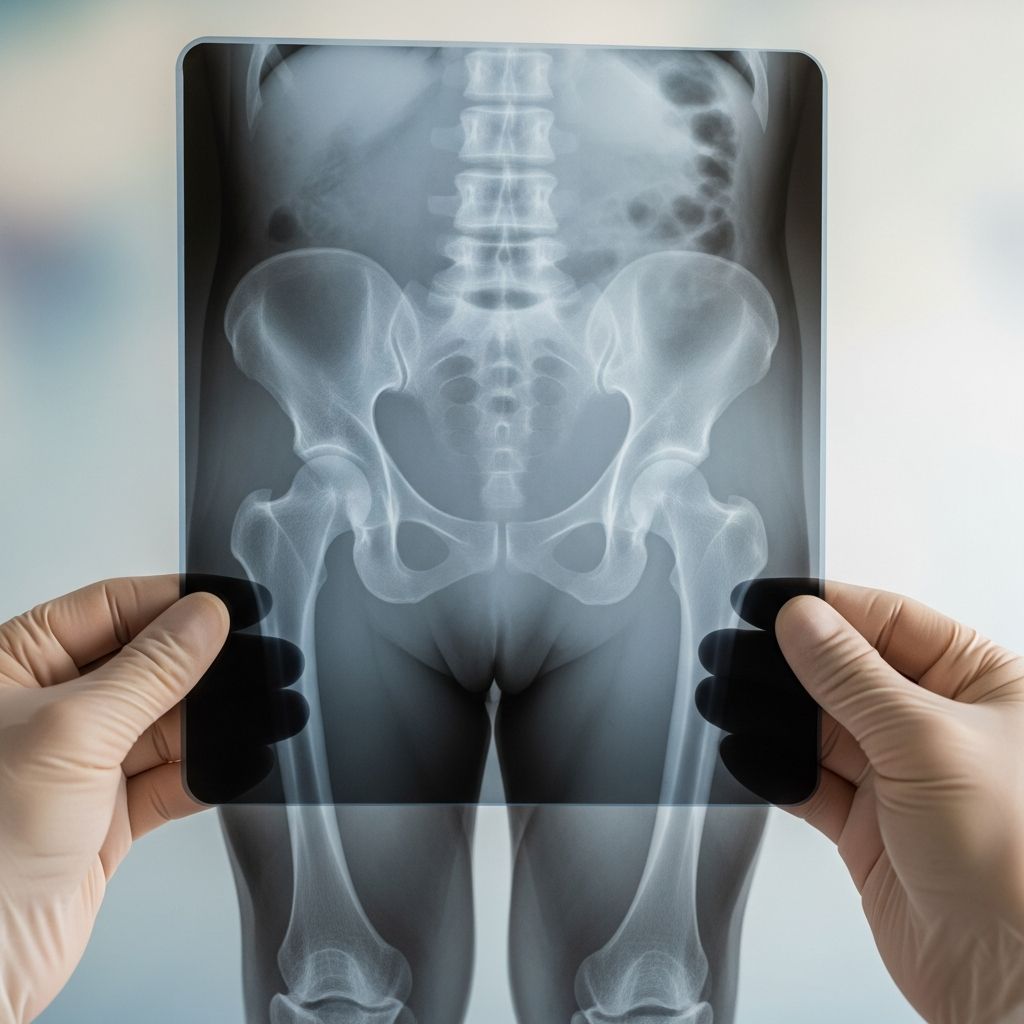
Understanding Pelvic Imaging in Children
Pelvic X-rays are among the most common diagnostic imaging procedures performed in pediatric medicine. These radiographic examinations allow healthcare providers to visualize the bones and structures within a child’s pelvic region, helping them identify potential health concerns, confirm diagnoses, and guide treatment decisions. For parents unfamiliar with this procedure, understanding what pelvic X-rays entail, why doctors recommend them, and what to expect can significantly reduce anxiety and help ensure a smooth imaging experience for their child.
The pelvic region contains numerous anatomical structures, including hip bones, the sacrum, and the coccyx. When a child experiences trauma, exhibits symptoms of hip or pelvic problems, or has a suspected underlying condition affecting these areas, a pelvic X-ray provides detailed visual information that other examination methods cannot offer. The procedure is quick, non-invasive, and involves minimal radiation exposure, making it an efficient diagnostic tool in pediatric care.
How Pelvic X-Ray Technology Functions
A pelvic X-ray works by directing a controlled beam of electromagnetic radiation through the child’s pelvic bones and surrounding tissue. The radiation passes through the body and is captured on specialized digital sensors or traditional radiographic film, creating a detailed black-and-white image. This imaging technology has evolved significantly over recent decades, with modern equipment producing clearer images while using substantially less radiation than earlier systems.
The bones appear white or light gray on the X-ray image because they are dense and absorb more radiation, while softer tissues and air-filled spaces appear darker. This contrast allows radiologists to distinguish between normal bone structure and abnormalities such as fractures, dislocations, or other pathological changes. The procedure produces no pain, no audible sounds, and no physical sensations for the child being examined.
Common Clinical Reasons for Recommending Pelvic X-Rays
Pediatricians and orthopedic specialists order pelvic X-rays for various clinical indications. Understanding these reasons can help parents recognize why their healthcare provider has recommended this imaging study.
- Trauma and Injury Assessment: Following accidents, falls, or injuries affecting the hip and pelvic region, X-rays help identify fractures, dislocations, or other bony injuries that may require intervention.
- Hip and Joint Concerns: Children presenting with persistent hip pain, groin discomfort, or limited mobility may require imaging to evaluate joint integrity and alignment.
- Developmental and Congenital Conditions: Certain developmental hip conditions that typically emerge during infancy and early childhood can be identified through pelvic radiography.
- Suspected Tumors or Lesions: When healthcare providers suspect abnormal bone growths or lesions within the pelvic region, X-rays provide initial diagnostic information.
- Evaluation of Chronic Conditions: Children with underlying bone diseases or metabolic disorders affecting skeletal development may need periodic pelvic X-rays to monitor their condition.
- Surgical Planning: When surgery involving the pelvis or hip is being considered, X-rays help surgeons understand the anatomical structures and plan their approach.
What Happens During a Pediatric Pelvic X-Ray Examination
Understanding the step-by-step process can help parents prepare their child and manage expectations. The typical pelvic X-ray examination follows a predictable sequence that most facilities have refined to minimize stress and ensure image quality.
Pre-Examination Preparation
Before entering the imaging room, a radiologic technologist will explain the procedure to both the parent and child in age-appropriate language. Parents typically receive a lead apron to wear during the examination, protecting them from radiation exposure while allowing them to remain present and support their child. Most imaging facilities permit at least one parent or guardian to stay in the room during the procedure, which helps anxious children remain calm.
Positioning and Image Acquisition
The child will lie on a flat X-ray table in a supine position, meaning on their back. The technologist will ensure the child’s body is properly positioned to obtain clear images. For optimal pelvic imaging, the child’s legs are typically positioned in slight internal rotation, a positioning technique that improves visualization of specific anatomical structures.
The technologist will take multiple images from different angles to provide comprehensive evaluation. The most common views include:
- Anteroposterior (AP) View: An image taken from the front with the child’s legs in a neutral or slightly internally rotated position. This view shows the overall pelvic structure and allows assessment of symmetry between sides.
- Lateral View: An image taken from the side, often with the child’s hips and knees slightly bent. This view provides depth perception and helps identify anterior or posterior displacement of structures.
- Specialized Views: Depending on clinical concerns, the technologist may obtain additional angled projections to evaluate specific regions such as the hip joint or sacroiliac joints.
During each exposure, the child must remain completely still for approximately 2-3 seconds. Movement during this brief interval can blur the image, potentially requiring repeat imaging. Technologists often ask older children to hold their breath during the exposure, as breathing movements can also compromise image quality. Younger children may require gentle physical guidance or reassurance to maintain stillness during this crucial moment.
Duration and Radiation Exposure
The entire pelvic X-ray examination, from initial positioning through final image verification, typically takes 10-15 minutes. However, the actual duration of radiation exposure is remarkably brief—usually less than one second total when all images are combined. This minimal exposure time is an important consideration when discussing radiation safety with parents.
Radiation Safety and Risk Considerations
Many parents express legitimate concerns about radiation exposure during medical imaging. Understanding both the risks and the safety measures in place can help parents make informed decisions and feel confident about their child’s care.
Modern X-ray equipment delivers very small amounts of radiation, and pelvic radiography is considered a low-radiation procedure. The actual biological risk from a single pelvic X-ray is extremely minimal. Healthcare providers follow the “ALARA” principle—As Low As Reasonably Achievable—when recommending imaging studies, meaning they only order X-rays when the diagnostic benefit clearly outweighs any potential radiation risk.
For most children undergoing a pelvic X-ray for legitimate clinical reasons, the diagnostic value far exceeds the minimal radiation exposure risk. However, special considerations apply in specific circumstances:
- Pregnancy Concerns: If a child might be pregnant, parents must inform the technologist before the procedure. While pelvic X-rays use very small radiation amounts, the radiologist and healthcare team will carefully weigh whether the imaging is truly necessary and will implement protective measures such as abdominal shielding.
- Recent Imaging: If a child has undergone other recent imaging studies involving radiation, inform the healthcare provider before scheduling a pelvic X-ray so cumulative exposure can be considered.
- Repeated Studies: When follow-up X-rays are needed to monitor a condition over time, healthcare providers will schedule these at appropriate intervals to minimize unnecessary cumulative exposure.
What Radiologists Look For in Pelvic X-Rays
After the technologist obtains the images and confirms they are clear and properly exposed, a radiologist—a physician with specialized training in interpreting medical images—reviews the films. Radiologists examine numerous details when evaluating pediatric pelvic X-rays, including bone alignment, structural integrity, joint positioning, and any abnormal features that might indicate pathology.
In children presenting with acute injury, radiologists assess for fractures involving various pelvic bones, including the pubic bones, iliac wings, sacrum, and coccyx. The radiologist determines fracture location, severity, and whether the fracture is stable or unstable. For children with hip-related complaints, radiologists evaluate the fit between the femoral head and the acetabular socket, assess for signs of avascular necrosis or other degenerative changes, and measure angles that help determine if surgical intervention might be necessary.
Results Interpretation and Follow-Up
Once the radiologist completes their evaluation, a detailed report is generated and sent to the child’s healthcare provider. The healthcare provider will review these findings with the family and explain what the X-rays show. Depending on the results, the healthcare provider may recommend additional diagnostic tests, such as advanced imaging with CT or MRI scans, or may proceed directly to treatment planning.
In some cases, follow-up X-rays may be recommended to monitor the child’s condition over time, track healing progress after an injury, or assess response to treatment. These follow-up studies help ensure the child’s condition is resolving as expected and allow the healthcare team to adjust treatment plans if necessary.
Preparing Your Child for a Pelvic X-Ray
Parents can take several steps to prepare their child for this procedure and ensure a positive experience:
- Explain the procedure using simple, reassuring language appropriate to your child’s age and comprehension level
- Emphasize that the procedure is painless and does not involve injections or special preparation
- Describe what the child will see and hear in the radiology room, such as the large machine and quiet beeping sounds
- Practice staying still for short periods before the appointment as a game or activity
- Bring a comfort item, such as a favorite stuffed animal, to the appointment if allowed by the facility
- Ask questions of the technologist and healthcare provider—most facilities are happy to address parent and child concerns
- Plan a calming post-appointment activity to help your child relax after the procedure
Frequently Asked Questions About Pediatric Pelvic X-Rays
How long does it take to get X-ray results?
Results timing varies by facility. In urgent situations, a radiologist may provide preliminary findings within hours. Routine studies typically receive formal written reports within 24-48 hours, which are then forwarded to your healthcare provider.
Is any special preparation required before a pelvic X-ray?
No special preparation is needed. Your child can eat, drink, and take regular medications normally. Simply wear comfortable, loose-fitting clothing without metal fasteners or decorations.
Can parents stay in the room during the X-ray?
Yes, parents are typically permitted to remain in the radiology room wearing a protective lead apron. Your presence can help keep your child calm and cooperative during the procedure.
Will my child feel any pain or discomfort?
The X-ray procedure itself causes no pain. However, if your child has an acute injury, positioning for certain views might cause mild discomfort. Inform the technologist if your child is in pain so they can work gently and take breaks if needed.
Are there any side effects from pelvic X-rays?
No side effects occur from the radiation exposure at the low levels used in diagnostic X-rays. Your child will not feel sick, dizzy, or experience any adverse reactions after the procedure.
What if my child cannot hold still during the X-ray?
Technologists are experienced working with children and use gentle guidance and reassurance. For very young or anxious children, the technologist may take extra time positioning the child or may require additional imaging attempts. In rare cases, sedation might be considered for children unable to cooperate.
Can pelvic X-rays detect all pelvic problems?
Pelvic X-rays are excellent for evaluating bone structure but have limitations. Advanced imaging such as ultrasound, CT, or MRI may be needed to evaluate soft tissues, organs, or internal structures in greater detail.
References
- X-Ray Exam: Pelvis — Rady Children’s Hospital System. https://www.rchsd.org/health-article/x-ray-exam-pelvis/
- Pediatric x-ray exam — Radiologyinfo.org (American College of Radiology). https://www.radiologyinfo.org/en/info/pediatric-xray
- Pelvis x-ray — UCSF Benioff Children’s Hospitals. https://www.ucsfbenioffchildrens.org/medical-tests/pelvis-x-ray
- Pelvis X-ray: Purpose, Procedure & Risks — Cleveland Clinic. https://my.clevelandclinic.org/health/diagnostics/23519-pelvis-x-ray