Magnetic Resonance Imaging (MRI) offers detailed views of a child’s internal structures using magnets and radio waves, making it a vital tool for diagnosing various conditions without radiation exposure.
Understanding the Power of Pediatric MRI
Pediatric MRI stands out as a cornerstone in modern diagnostic medicine for children. This imaging technique harnesses a strong magnetic field combined with radiofrequency pulses to generate high-resolution images of soft tissues, organs, bones, and blood vessels. Unlike traditional X-rays or CT scans, it avoids ionizing radiation, which is particularly beneficial for growing bodies.
The process begins with hydrogen atoms in the body aligning to the magnetic field. Radio waves then disrupt this alignment temporarily, and as atoms realign, they emit signals captured by the scanner to form cross-sectional images. These can be reconstructed into 3D models, providing physicians with unparalleled insights into complex anatomical areas.
For children, MRI is invaluable because it excels at visualizing areas obscured by bone, such as the brain and spinal cord. It helps detect subtle abnormalities that other modalities might miss, ensuring precise diagnoses and tailored treatment plans.
Why Doctors Recommend MRI for Kids
Physicians order pediatric MRIs to investigate a broad spectrum of health issues. In neurology, it’s used to identify brain tumors, cysts, inflammation from infections like encephalitis, stroke damage, or congenital malformations. It also evaluates chronic conditions such as multiple sclerosis or pituitary gland disorders, and assesses developmental delays or seizure causes.
For the spine, MRI reveals herniated discs, nerve compression, or spinal cord injuries. In the abdomen and pelvis, it pinpoints sources of pain like appendicitis, monitors infections, inflammatory diseases, or cancer treatment progress. Orthopedic applications include diagnosing sports injuries, joint infections, bone cancers, or fractures in children.
| Body Region | Common Uses |
|---|---|
| Brain & Spine | Tumors, injuries, infections, developmental issues |
| Abdomen/Pelvis | Pain causes, trauma, cancer monitoring |
| Bones & Joints | Injuries, infections, tumors, marrow diseases |
This table summarizes key applications, highlighting MRI’s versatility across pediatric care.
Step-by-Step: What Happens During the Scan
A child’s MRI is typically an outpatient procedure lasting 25 to 90 minutes, depending on the scanned area and sequences required. Upon arrival, staff screen for metal implants, pacemakers, or claustrophobia risks, as the magnet can interact with ferromagnetic materials.
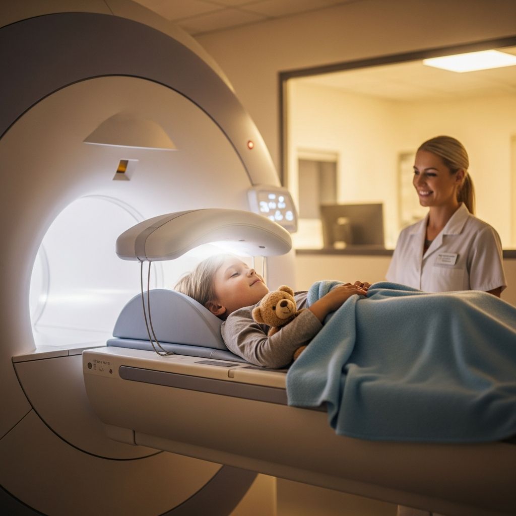
The child changes into a gown and removes all metal objects like jewelry or hairpins. They’re positioned on a padded table with straps for stability. Coils—special antennas—are placed near the imaging area to enhance signal quality. The table slides into the scanner’s tunnel, which is open-ended in many pediatric units.
The machine produces rhythmic banging noises as gradients activate for different image contrasts. Multiple sequences follow, each lasting 2-10 minutes, requiring the child to remain still. Parents or caregivers often stay nearby, communicating via intercom, and children may watch videos through mirrors or wear headphones for music.
If contrast is needed—gadolinium-based for better vessel or tumor visibility—an IV line is inserted beforehand. A saline flush keeps it open, and contrast is injected mid-scan, potentially causing a cool sensation or metallic taste. Post-scan, images are reviewed; additional sequences may be needed.
- Preparation: Screening questionnaire and gown change.
- Positioning: Comfortable setup with supports.
- Scanning: Noisy sequences with breaks to relax.
- Contrast (if used): Quick IV injection.
- Completion: IV removal and image check.
Preparing Your Child for a Successful MRI
Preparation eases anxiety and ensures quality images. For infants and toddlers under sedation guidelines, fasting may be required; consult your doctor. Older children benefit from age-appropriate explanations: compare the scanner to a spaceship or amusement ride, emphasizing it’s painless.
Practice sessions help: mimic the table with a blanket tunnel at home, play MRI sound samples from hospital apps, and role-play staying still. Tour the facility if offered, or watch child-friendly videos showing the process.
Pack comforts: favorite blanket, stuffed animal (metal-free), headphones playlist. Sedation is an option for very young or anxious kids, using oral meds or anesthesia monitored by specialists. Discuss with your provider.
On scan day, arrive early. Staff use child life specialists to distract with stories or gadgets during IV placement.
Safety First: Risks and Precautions
MRI is safe for all ages, including fetuses, with no known long-term effects from the magnetic field or radio waves. Primary concerns are implants: cochlear devices, aneurysm clips, or certain tattoos may heat up or move. A detailed safety checklist mitigates this.
Contrast agents rarely cause allergic reactions (less than 1 in 10,000), but those with kidney issues need monitoring. Claustrophobia affects some; open MRI designs or mild sedatives help. Noise is loud (up to 100 decibels), but ear protection is standard.
Post-contrast, hydrate well. Bruising at IV sites is minor and temporary.
Advanced Techniques in Pediatric Imaging
Beyond standard MRI, functional MRI (fMRI) maps brain activity, useful for pre-surgical planning. MR spectroscopy analyzes tissue chemistry for tumor typing. Diffusion tensor imaging tracks nerve fibers.
Pediatric centers employ motion-correction software and faster sequences to accommodate wiggly kids, reducing sedation needs.
After the Scan: Next Steps and Care
Most children resume normal activities immediately. A radiologist analyzes images, sending a report to your doctor within days to a week due to volume. Follow-up discusses findings and plans.
Monitor for delayed contrast reactions (rash, nausea)—rare. Praise your child’s bravery to build resilience.
Frequently Asked Questions (FAQs)
Is MRI painful for children?
No, it’s painless. The only discomfort might be from IV insertion, which is brief.
How long does a kid’s MRI take?
Typically 25-60 minutes, varying by body part and complexity.
Can parents stay with their child?
Yes, in the room or nearby, unless sedation requires separation.
Does MRI use radiation?
No, it uses magnets and radio waves only.
What if my child is scared of loud noises?
Headphones and music block sounds effectively.
Is sedation always needed?
Not for cooperative older kids; preparation techniques often suffice.
This guide equips parents to navigate the MRI process confidently, prioritizing comfort and clear results.
References
- Pediatric MRI — RadiologyInfo.org. 2023-10-01. https://www.radiologyinfo.org/en/info/pediatric-mri
- MRI scan — Royal Children’s Hospital Melbourne. 2024-05-15. https://www.rch.org.au/kidsinfo/fact_sheets/MRI_scan/
- Magnetic Resonance Imaging (MRI) — Nemours KidsHealth. 2024-02-20. https://kidshealth.org/en/parents/mri.html
- Preparing for Your Child’s MRI — Children’s Nebraska. 2024-08-10. https://www.childrensnebraska.org/radiology-and-imaging-services/preparing-for-your-childs-mri
- Pediatric MRI: What to Expect — Children’s Hospital Colorado (YouTube). 2024-08-22. https://www.youtube.com/watch?v=vTFjGDdSFDY