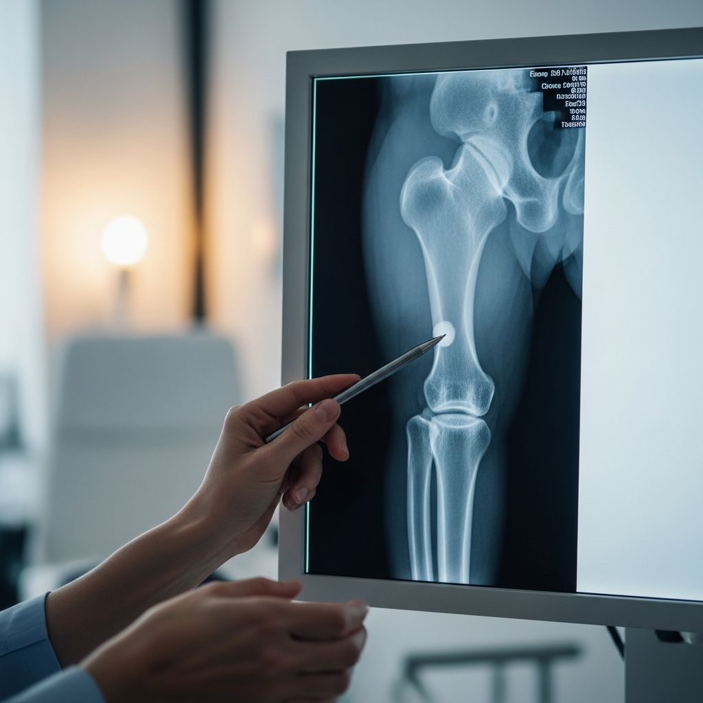
When a child hurts a shin, ankle, or lower leg, a doctor may recommend an X-ray to look inside the bones and joints without surgery. Understanding why this test is needed and what happens during the scan can make the experience calmer for both children and parents.
Why Doctors Order Lower Leg X-Rays
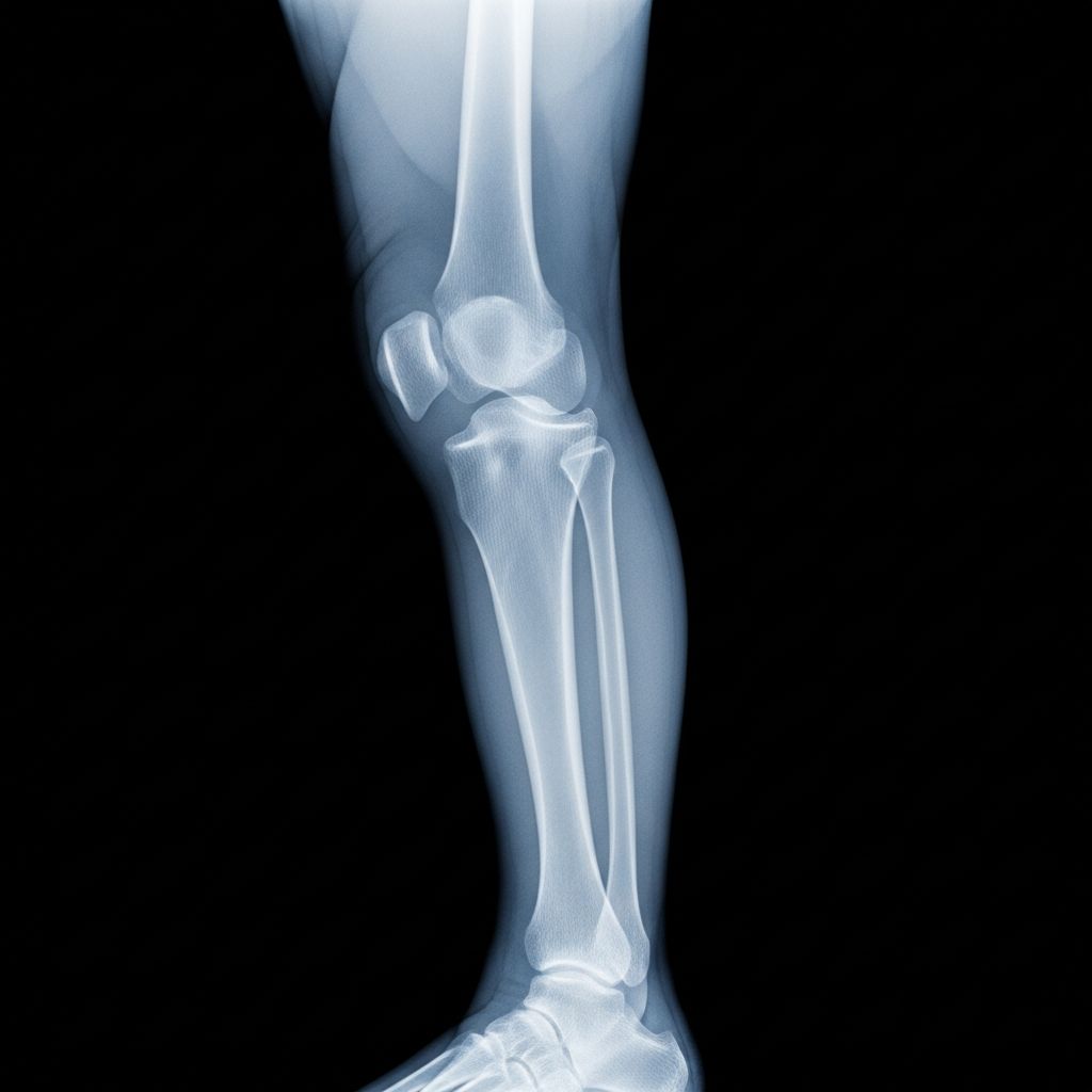
A lower leg X-ray is a quick imaging test that creates pictures of the bones between the knee and the ankle, mainly the tibia (shin bone) and fibula (the smaller bone beside it). Doctors use these images to check for injuries, track healing, and look for signs of disease that may not be obvious from the outside.
Common Reasons a Child Might Need One
- After a fall, sports injury, or accident to check for fractures or bone bruises.
- Persistent pain, limping, or swelling in the lower leg without a clear cause.
- Visible deformity or bending of the leg after trauma.
- Infection concerns, such as fever plus bone pain, to look for bone infection.
- Follow-up after a fracture has been put in a cast or splint to be sure the bones are lined up well and healing correctly.
What Exactly Does the X-Ray Show?
An X-ray uses a small amount of radiation that passes through the body to create detailed black-and-white images. On these images, dense structures like bone appear white or light gray, while softer tissues appear darker, helping doctors tell what is normal and what is not.
Bones and Structures in the Lower Leg
- Tibia: The bigger, weight-bearing shin bone in the front of the lower leg.
- Fibula: The thinner bone along the outside of the lower leg that helps stabilize the ankle.
- Knee joint area: The lower end of the thigh bone and the upper ends of the tibia and fibula.
- Ankle joint area: The lower ends of the tibia and fibula and the upper ankle bones.
- Growth plates: Special areas near the ends of long bones where a child’s bones lengthen as they grow.
Is a Lower Leg X-Ray Safe for My Child?
X-rays do use radiation, but modern equipment is designed to use the smallest amount possible while still producing clear images. For most children, the radiation from a single leg X-ray is very low and is considered a safe tool when used for a good medical reason.
Imaging teams follow strict safety rules to protect children, including shielding nearby body areas with lead coverings when possible and focusing the beam only where it is needed. Parents who are pregnant or might be pregnant are usually asked to step back during exposure or wear protective shielding as well.
Preparing Your Child: Before the Appointment
Good preparation can turn a scary, unknown test into a predictable experience for a child. A few simple steps can reduce anxiety and help the appointment go faster and more smoothly.
How Parents Can Get Ready
- Bring your child’s health card, imaging order, and any relevant medical notes.
- Have a list of medications, allergies, and recent illnesses ready in case staff ask.
- Dress your child in comfortable, loose clothing that can be rolled up above the knee.
- Leave jewelry and metal accessories at home if possible, especially ankle bracelets or metal decorations on pants.
- Arrive a little early to allow time for parking, check-in, and forms.
Explaining the Test to Your Child
Children cope better when they know what to expect in age-appropriate language. Keep explanations short, honest, and calm, avoiding words that may sound frightening.
- Describe the machine as a special camera that takes pictures of bones.
- Explain that the test does not cut, poke, or go inside the body.
- Let them know they will need to hold still like a statue for a few seconds at a time.
- Offer reassurance that you or another trusted adult will be nearby.
Step-by-Step: What Happens During a Lower Leg X-Ray
The entire visit is usually short, often less than half an hour, with the actual X-ray exposures taking just a few seconds each. The process is handled by a trained radiologic technologist who specializes in capturing clear images while keeping children safe and as comfortable as possible.
From Check-In to Positioning
- Check-in: You arrive at the imaging center or hospital, complete any necessary forms, and wait to be called.
- Safety questions: Staff may ask if your child could be pregnant (for older teens), has metal implants, or has had recent imaging tests.
- Changing clothes, if needed: Your child may be asked to wear a gown if clothing has metal snaps, zippers, or thick fabric over the lower leg.
- Entering the X-ray room: The technologist explains what will happen in simple terms and answers your questions.
- Positioning on the table: Your child usually lies on a padded table, with the injured leg gently placed in the right position.
During the Actual X-Ray
To see the bones clearly, the technologist usually takes at least two pictures from different angles, often front and side views. Cushions, foam blocks, or straps may be used to keep the leg steady so the images do not come out blurry.
- The technologist moves the X-ray camera over the leg and places a detector or plate under or beside it.
- Your child is asked to keep very still and may be told to hold their breath for just a moment.
- The technologist steps behind a protective screen or into a nearby area to activate the machine.
- A soft clicking sound may be heard when the picture is taken, but your child will not feel the X-ray.
- This may be repeated for another angle, such as turning the leg slightly or rolling onto the side.
How to Keep Your Child Calm and Comfortable
Emotional comfort is just as important as physical comfort, especially for younger children or those with previous medical fears. Parents can play a huge role in turning the test into a manageable experience.
Comfort Strategies That Help
- Bring a favorite small toy, stuffed animal, or blanket for your child to hold.
- Practice “freeze” games at home so staying still feels familiar and even fun.
- Use calm, steady breathing together if your child looks nervous.
- Ask the technologist if you may stand near your child (often wearing a lead apron) for reassurance.
- Offer praise and small rewards afterward, such as a sticker or special activity.
What Happens After the X-Ray?
Once the images are taken, your child can usually get dressed and leave right away unless more tests are needed. There is no recovery time because the test is painless and noninvasive.
A doctor trained in interpreting medical images, often called a radiologist, reviews the pictures and writes a report. Your child’s main doctor then uses that report, along with the physical exam and symptoms, to decide on the next steps in care.
Possible Next Steps
- No fracture found: The doctor may recommend rest, ice, and follow-up if symptoms persist.
- Minor fracture or hairline crack: A splint, boot, or cast may be used to keep the bone stable while it heals.
- More serious fracture: A referral to an orthopedic specialist may be needed to discuss treatment options.
- Signs of infection or other conditions: Additional blood tests or imaging, such as MRI or ultrasound, may be ordered.
Typical Lower Leg X-Ray Timeline
Every clinic is a little different, but most follow a similar pattern from arrival to results. The table below gives a general idea of how long each part of the visit may take when things run smoothly.
| Stage | What Happens | Estimated Time |
|---|---|---|
| Check-In | Registration, forms, insurance details if needed | 5–10 minutes |
| Preparation | Questions about health, changing into a gown if required | 5–10 minutes |
| Positioning | Moving your child onto the table, placing the leg correctly | 5–10 minutes |
| Imaging | Taking 2–3 views from different angles | 5–10 minutes |
| Post-Exam | Dressing, leaving the imaging area | 5–10 minutes |
How to Read Your Child’s X-Ray Report (In Simple Terms)
Medical reports are usually written for healthcare professionals, but a few key words can help parents make sense of the results. Always ask your child’s doctor to walk through the report and answer any questions in plain language.
Common Terms You Might See
- Normal alignment: The bones are in the correct position relative to each other.
- No acute fracture: No new break in the bone is seen.
- Hairline or nondisplaced fracture: A thin crack is present, but the bone pieces have not shifted out of place.
- Displaced fracture: Bone pieces have moved out of alignment.
- Soft tissue swelling: The muscles and skin around the bone look swollen, often because of injury.
- Growth plate involvement: The injury affects the growth area, which may require closer follow-up.
Helping Your Child After the Appointment
Most children feel completely normal after a lower leg X-ray and can return to regular activities unless the doctor has advised rest because of an injury. The key tasks for parents are watching symptoms and following any care instructions given by the healthcare team.
Post-Visit Care Tips
- Keep the leg elevated if swelling or pain is present.
- Use ice packs over a cloth for short intervals if recommended by the doctor.
- Follow dosing instructions carefully for any pain medicine prescribed or suggested.
- Encourage gentle rest until you know whether a fracture or serious injury is present.
- Call the doctor if pain suddenly worsens, fever develops, or the leg changes color or temperature.
Frequently Asked Questions About Lower Leg X-Rays in Children
Does a lower leg X-ray hurt?
The X-ray itself is painless because the machine does not touch or go inside the body. The only discomfort usually comes from having to hold the leg in certain positions, which can be slightly uncomfortable if the leg is already sore.
How long does it take to get results?
In urgent situations, such as a suspected serious fracture, a preliminary reading may be available within minutes. For less urgent cases, the report may take a few hours to a day, depending on the clinic and time of day, and results are typically shared by your child’s main doctor.
Can I stay with my child during the X-ray?
In many facilities, one parent or caregiver is allowed to stay in the room during the test, especially for younger children. You may be asked to wear a lead apron and stand in a specific location to limit your own exposure to radiation.
What if my child cannot stay still?
Technologists who work with children every day use creative techniques, such as games or counting, to help them hold still for a few seconds at a time. In rare cases, if a child cannot cooperate and the images are essential, the doctor might discuss other options, but this is uncommon for a simple leg X-ray.
Is one X-ray enough if my child has frequent injuries?
Each X-ray adds a small amount of radiation, so doctors weigh the benefits of getting clear information against the small risk from exposure. If your child has repeated injuries, the doctor may suggest preventive strategies, physical therapy, or other types of imaging when appropriate.
Could another test replace the X-ray?
For bone injuries, X-rays are often the first and most efficient choice because they are fast, available, and give clear pictures of bones. Ultrasound, CT, or MRI may be used in special situations, but these tests can be more time-consuming, more expensive, or require special equipment and preparation.
Key Takeaways for Parents
- A lower leg X-ray is a quick, safe, and effective way for doctors to check your child’s tibia, fibula, and nearby joints.
- The test uses a very small amount of radiation and does not hurt, though injured legs may be sore when positioned.
- Preparing your child with simple explanations and comfort items can greatly reduce stress.
- Results from the X-ray help guide decisions about casts, splints, rest, or other treatments.
- Never hesitate to ask the healthcare team to explain each step and clarify the results in everyday language.