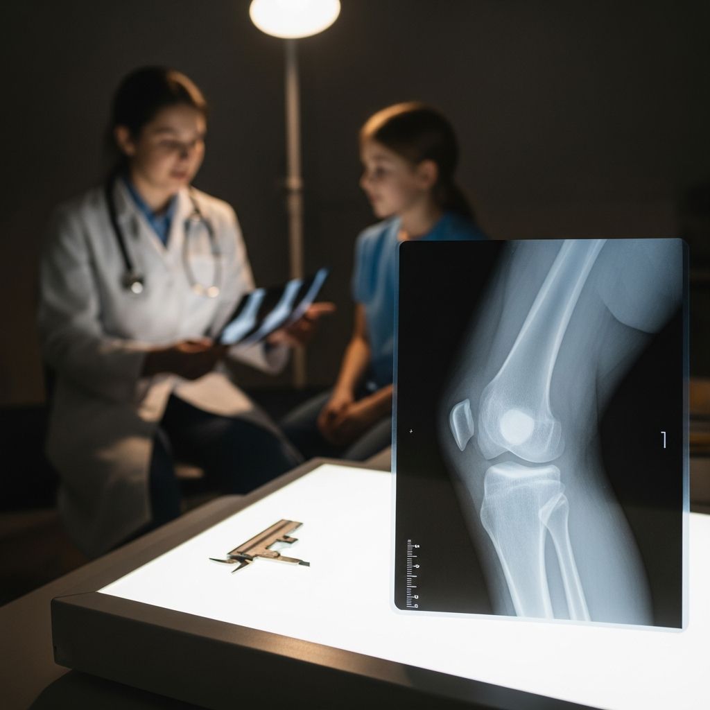
Why Doctors Recommend Knee X-Rays for Kids
When a child injures their knee or experiences persistent pain, swelling, or difficulty walking, a knee X-ray is often one of the first diagnostic tools a pediatrician or orthopedic specialist will recommend. Unlike soft tissue imaging such as MRI, X-rays are excellent at visualizing bones, making them ideal for detecting fractures, dislocations, growth plate injuries, and certain developmental changes around the knee joint.
Common reasons a doctor might order a knee X-ray include:
- A fall, sports injury, or direct blow to the knee
- Swelling, bruising, or visible deformity after trauma
- Refusal or inability to bear weight on the leg
- Pain that worsens with activity or persists beyond a few days
- Concerns about alignment, such as knock-knees or bowlegs
- Monitoring known conditions like Osgood-Schlatter disease or patellar instability
While X-rays don’t show ligaments, tendons, or cartilage in detail, they provide crucial information about the bony structures that support the knee, helping doctors decide whether further imaging (like MRI or ultrasound) is needed.
How a Pediatric Knee X-Ray Is Performed
A knee X-ray is a quick, non-invasive procedure that typically takes place in a doctor’s office, urgent care center, or hospital radiology department. The process is designed to be as comfortable and efficient as possible, especially for children who may feel anxious about medical tests.
What Happens During the Exam
A radiologic technologist, trained in pediatric imaging, will guide your child through the steps:
- Your child will be asked to sit or lie down on an X-ray table, depending on their age and comfort level.
- The technologist will gently position the knee in specific ways to capture different views. Common views include:
- Anteroposterior (AP) view: The child lies on their back with the knee straight and the foot pointing up.
- Lateral view: The child lies on their side with the knee slightly bent, allowing a side profile of the joint.
- Patellar (sunrise or tangential) view: The knee is bent and the X-ray beam is angled to show the kneecap and its relationship to the femur.
- The technologist will step behind a protective barrier or into another room to activate the X-ray machine.
- Your child will need to stay still for a few seconds while each image is taken. Movement can blur the picture, which might require repeating the view.
What Parents Can Do to Help
Children often pick up on parental anxiety, so staying calm and reassuring can make a big difference:
- Explain the process in simple terms: “The machine will take pictures of your knee, like a camera, but it doesn’t hurt.”
- Bring a favorite toy, book, or tablet to help your child stay still and distracted.
- Ask if you can stay in the room; many facilities allow one parent to remain nearby.
- Practice “holding still like a statue” at home before the appointment.
What a Knee X-Ray Can and Cannot Show
Understanding the capabilities and limitations of a knee X-ray helps parents interpret what the doctor is looking for and why additional tests might be ordered.
What X-Rays Reveal
Knee X-rays are excellent for evaluating:
- Fractures: Breaks in the femur (thigh bone), tibia (shin bone), fibula, or patella (kneecap).
- Growth plate injuries: In children, the growth plates (physis) near the ends of long bones are vulnerable to injury and appear as dark lines on X-rays.
- Dislocations: When the kneecap or other bones are out of their normal position.
- Bone alignment: Whether the leg bones are properly aligned, which is important in conditions like knock-knees or bowlegs.
- Joint space: Narrowing or widening of the space between bones can suggest arthritis, injury, or other joint problems.
- Developmental changes: Conditions like Osgood-Schlatter disease (irregularity at the tibial tubercle) or Sinding-Larsen-Johansson syndrome (changes at the bottom of the kneecap) often show characteristic findings on X-ray.
What X-Rays Do Not Show Well
Because X-rays primarily image dense structures, they are less effective at visualizing:
- Ligaments (like the ACL or MCL)
- Tendons (such as the patellar or quadriceps tendon)
- Meniscus tears
- Cartilage damage
- Early soft tissue swelling or inflammation
When soft tissue injury is suspected, doctors may recommend an MRI, ultrasound, or sometimes a CT scan to get a more complete picture of the knee’s internal structures.
Interpreting the Results: What the Radiologist Looks For
After the X-ray is taken, a radiologist (a doctor specialized in medical imaging) examines the images and prepares a report for your child’s physician. The radiologist evaluates several key aspects of the knee:
Key Areas Checked on a Knee X-Ray
| Area | What Is Assessed |
|---|---|
| Femur and tibia | Fractures, alignment, growth plate appearance |
| Patella (kneecap) | Position, fractures, alignment with femur |
| Tibial tubercle | Irregularity or fragmentation in active children (Osgood-Schlatter) |
| Joint space | Width between bones, signs of narrowing or effusion |
| Soft tissue | Swelling, fat pad displacement, or large effusions |
Common Findings and What They Might Mean
- Normal X-ray: No fractures, dislocations, or significant alignment issues. Pain may be due to soft tissue injury, overuse, or minor sprain.
- Fracture: A break in the bone, which may be treated with casting, bracing, or, in some cases, surgery.
- Growth plate injury: Requires careful management to avoid long-term growth problems; treatment depends on the type and severity.
- Patellar malalignment: The kneecap sits too far to the side, which can contribute to pain and instability.
- Effusion (fluid in the joint): Often seen as a bulge in the soft tissues; can indicate trauma, infection, or inflammatory conditions.
- Developmental changes: Findings like those in Osgood-Schlatter or Sinding-Larsen-Johansson are common in active children and usually resolve with rest and activity modification.
Safety and Radiation: What Parents Should Know
Many parents worry about radiation exposure when their child needs an X-ray. It’s important to understand that modern X-ray equipment uses very low doses of radiation, and the benefits of an accurate diagnosis usually far outweigh the small risks.
Radiation Dose in Pediatric Knee X-Rays
- A typical knee X-ray exposes a child to a very small amount of radiation, comparable to the natural background radiation received over a few days.
- Technologists use the lowest possible dose needed to get a clear image, following the ALARA principle (As Low As Reasonably Achievable).
- Lead shields (aprons or collars) are often used to protect sensitive areas like the abdomen and thyroid, especially in younger children.
When X-Rays Are Avoided or Modified
In certain situations, doctors may adjust how or whether an X-ray is done:
- If a child is pregnant (in older adolescents), the healthcare team will carefully weigh the need for imaging and take extra precautions.
- For very young children or those with special needs, sedation is rarely needed for a simple knee X-ray, but staff are trained to work with children of all ages and abilities.
- When possible, doctors use clinical decision rules (like the Ottawa Knee Rules in older children) to avoid unnecessary X-rays in low-risk cases.
Preparing Your Child for a Knee X-Ray
Good preparation can reduce anxiety and help the exam go smoothly. Here’s how to get ready:
Before the Appointment
- Explain what will happen in age-appropriate language.
- Let your child wear comfortable, loose-fitting clothes; they may need to change into a gown.
- Remove metal objects (belts, jewelry, some clothing with zippers or snaps) from the area being imaged.
- Bring any previous imaging or medical records related to the knee, if available.
During the Appointment
- Stay calm and reassuring; your child will take cues from you.
- Encourage your child to follow the technologist’s instructions about staying still.
- Ask questions if you’re unsure about the procedure or why it’s needed.
After the X-Ray
- There are no restrictions after a knee X-ray; your child can resume normal activities unless otherwise instructed.
- Results are typically reviewed by the doctor within a day or two, depending on the setting.
- Follow up with your child’s healthcare provider to discuss the findings and next steps.
When Further Testing Might Be Needed
A normal X-ray doesn’t always mean everything is fine. If symptoms persist or the doctor suspects a soft tissue injury, additional tests may be recommended:
- MRI: Best for detailed images of ligaments, tendons, cartilage, and bone marrow.
- Ultrasound: Useful for evaluating tendons, fluid collections, and some growth-related conditions.
- CT scan: Sometimes used for complex fractures or pre-surgical planning, though it involves higher radiation than X-ray.
- Follow-up X-rays: May be ordered to monitor healing of a fracture or growth plate injury over time.
Frequently Asked Questions
Q: Does a knee X-ray hurt?
A: No, the X-ray itself is painless. Your child might feel some discomfort if the knee is already injured when it’s positioned, but the technologist will be as gentle as possible.
Q: How long does a knee X-ray take?
A: The actual imaging usually takes only a few minutes. The entire appointment, including check-in and positioning, may last 15–30 minutes.
Q: Can my child eat or drink before the X-ray?
A: Yes, there are no dietary restrictions for a standard knee X-ray. Your child can eat and drink normally.
Q: How soon will we get the results?
A: In urgent settings (like the ER), results may be discussed within hours. In clinics, it often takes 1–2 days for the radiologist’s report to be available to your child’s doctor.
Q: Are there any side effects from the radiation?
A: The radiation dose from a knee X-ray is very low and not associated with immediate side effects. The long-term risk is extremely small, especially when the test is medically necessary.
Q: What if the X-ray is normal but my child still has pain?
A: A normal X-ray means there’s no obvious fracture or dislocation, but soft tissue injuries (sprains, tendinitis, overuse) can still cause significant pain. Your doctor may recommend rest, physical therapy, or further imaging if symptoms don’t improve.
References
- ACR Appropriateness Criteria®: Suspected Knee Trauma – Child — American College of Radiology. 2023. https://www.acr.org/Clinical-Resources/ACR-Appropriateness-Criteria
- Knee Radiography in Children: Technique and Interpretation — Radiopaedia.org. 2024. https://radiopaedia.org/articles/knee-radiograph
- Pediatric Knee Injuries: Evaluation and Management — UpToDate. 2024. https://www.uptodate.com/contents/pediatric-knee-injuries-evaluation-and-management