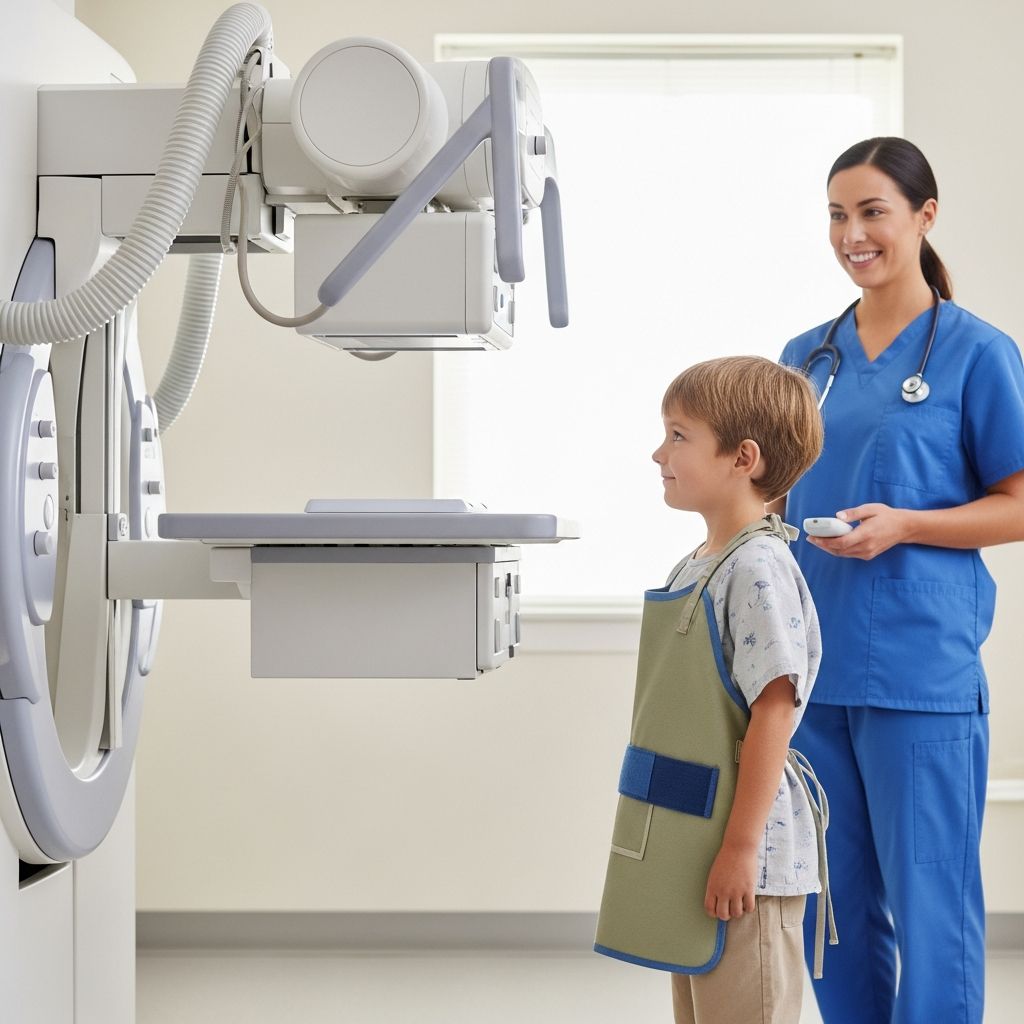
Chest X-rays remain one of the most common and vital diagnostic tools in pediatric care, offering quick snapshots of a child’s lungs, heart, and surrounding structures. For parents, understanding this simple yet powerful procedure can reduce anxiety and ensure a smoother experience for their little ones.
Why Doctors Recommend Chest X-Rays for Kids
Pediatricians often turn to chest X-rays when investigating respiratory issues or other thoracic concerns in children. These images help visualize the lungs, heart, ribs, and diaphragm, revealing conditions that might not be apparent through physical exams alone. Common reasons include persistent coughs, difficulty breathing, fevers of unknown origin, or chest injuries from play or accidents.
Infections like pneumonia show up as areas of consolidation in the lung fields, while conditions such as asthma exacerbations or foreign body aspirations can also be assessed. Heart-related issues, including enlarged cardiac silhouettes suggestive of congenital defects, are another frequent indication. Early detection through these radiographs can lead to timely interventions, preventing complications.
For infants and toddlers, chest X-rays are particularly useful in neonatal intensive care units to monitor lung maturity or device placements like endotracheal tubes. In older children, they aid in diagnosing sports-related injuries or monitoring chronic conditions like cystic fibrosis.
Understanding the Science Behind Chest X-Rays
X-rays work by emitting a small burst of ionizing radiation that passes through the body, absorbed differently by various tissues. Air-filled lungs appear dark (radiolucent), while denser bones and heart tissue show as lighter shades (radiopaque). This contrast creates a detailed 2D image of the chest cavity.
Modern digital systems capture these images instantly, allowing radiologists to zoom, adjust contrast, and detect subtle abnormalities like small nodules or fluid accumulations. For children, low-dose protocols minimize exposure while maintaining diagnostic quality.
| Structure | Appearance | Common Abnormalities |
|---|---|---|
| Lungs | Dark fields with branching vessels | Consolidation, nodules, hyperinflation |
| Heart | Central white silhouette | Enlargement, border irregularities |
| Diaphragm | Dome-shaped below lungs | Elevation, free air below |
| Ribs & Spine | Linear white densities | Fractures, alignment issues |
Preparation Steps for Your Child’s Chest X-Ray
Preparing a child for a chest X-ray is straightforward, but a few steps can make it less intimidating. Explain the process in age-appropriate terms: it’s like standing still for a quick photo that helps the doctor see inside their chest to make them feel better.
- Remove all metal objects: Jewelry, belts, zippers, or buttons can obscure images. Dress your child in loose clothing without snaps.
- Check for pregnancy: Though rare in kids, inform staff if applicable, as radiation should be avoided.
- Practice deep breaths: Teach older children to inhale deeply and hold when instructed.
- Bring comfort items: A favorite toy or blanket can soothe toddlers during waits.
- Fast if required: Rarely needed, but confirm for any contrast studies.
For infants, feeding right before might help them stay calm, but avoid if sedation is planned. Hospitals often have child life specialists to demonstrate the machine and ease fears.
Step-by-Step: What Happens During the Exam
The procedure typically takes just minutes in a radiology suite. Your child stands or sits between an X-ray tube and a digital detector plate. Technologists, trained in pediatrics, ensure proper positioning for clear PA (posteroanterior) and lateral views.
- Front View (PA): Child faces the plate, shoulders rolled forward, arms at sides or raised. Take a deep breath and hold— this expands lungs for better visualization.
- Side View (Lateral): Turn sideways, arms up, chest against the plate. Hold breath again.
- For young or uncooperative kids: May use a seated position, immobilization devices, or parental holding with lead shielding.
- Images are reviewed immediately; repeats are rare with digital tech.
Parents can often stay in the room, wearing lead aprons to shield from scatter radiation. No pain involved—just stay still!
Radiation Safety: Protecting Your Child
Concerns about radiation are valid, but pediatric chest X-rays use extremely low doses—equivalent to a few days of natural background radiation. Advanced equipment like pulsed fluoroscopy and collimation (limiting beam size) further reduces exposure.
Benefits always outweigh risks for medically necessary scans. The ALARA principle (As Low As Reasonably Achievable) guides protocols. For context:
- Single chest X-ray: ~0.01-0.1 mSv
- Annual background radiation: ~3 mSv
- Cross-country flight: ~0.03 mSv
Pregnant mothers should discuss alternatives like ultrasound, but for kids, risks of undiagnosed conditions far exceed radiation effects.
Decoding the Results: What Radiologists Look For
A pediatric radiologist analyzes the images systematically. They start with technical quality, then scan from periphery inward: soft tissues, bones, lungs, heart, mediastinum.
Normal findings: Clear lung fields, normal heart size (cardiothoracic ratio <0.5), sharp diaphragm domes, no masses. Abnormalities might include:
- Opacity: Pneumonia, aspiration.
- Hyperlucency: Pneumothorax, emphysema.
- Enlarged heart: Cardiomyopathy, effusion.
- Device checks: Tube positions in ICU cases.
Results go to the ordering doctor promptly, often same day. Follow-ups like CT or MRI if needed.
When Follow-Up Imaging Might Be Needed
Chest X-rays often serve as a starting point. If findings suggest complexity, such as masses or unclear infections, further tests follow:
- Ultrasound: For pleural effusions or heart function.
- CT scan: Detailed 3D views, higher radiation but justified when necessary.
- MRI: No radiation, best for soft tissues.
- Echo: Heart-specific.
Comparing with prior images tracks changes, crucial for chronic conditions.
Common Myths and Facts About Pediatric Chest X-Rays
| Myth | Fact |
|---|---|
| X-rays cause cancer in kids. | Risk is negligible; benefits dominate. |
| Kids must be sedated. | Rarely; most cooperate with coaching. |
| It’s painful. | No—quick and painless. |
| Results take weeks. | Usually immediate review. |
Frequently Asked Questions (FAQs)
Is a chest X-ray safe for my baby?
Yes, doses are minimized for infants, using shielding and brief exposures. Safer than missing a diagnosis.
How long does it take?
5-10 minutes total, with actual exposures under 1 second.
Can I be with my child?
Typically yes, with protective gear.
What if my child can’t stay still?
Technicians use gentle restraints or distractions; sedation only if essential.
Will insurance cover it?
Usually, as it’s standard diagnostic.
Empowering Parents: Next Steps After the X-Ray
Discuss results with your pediatrician. Ask about lifestyle changes, medications, or specialist referrals. Routine follow-ups ensure ongoing health. Knowledge demystifies the process, fostering confidence in your child’s care journey.
By understanding chest X-rays, parents become active partners in pediatric health, ensuring quick resolutions to common childhood ailments.
References
- Chest X-Ray – Basic Interpretation — The Radiology Assistant. 2023. https://radiologyassistant.nl/chest/chest-x-ray/basic-interpretation
- Chest X-rays — Mayo Clinic. 2024-05-12. https://www.mayoclinic.org/tests-procedures/chest-x-rays/about/pac-20393494
- Chest X-ray (Radiography) — RadiologyInfo.org (RSNA). 2024. https://www.radiologyinfo.org/en/info/chestrad
- Chest X-ray — Quirónsalud. 2023. https://www.quironsalud.com/en/diagnostic-tests/chest-x-ray
- Chest X-ray Interpretation: Detecting Devices and Device-Related Complications — PMC (NCBI). 2023-02-20. https://pmc.ncbi.nlm.nih.gov/articles/PMC9954842/