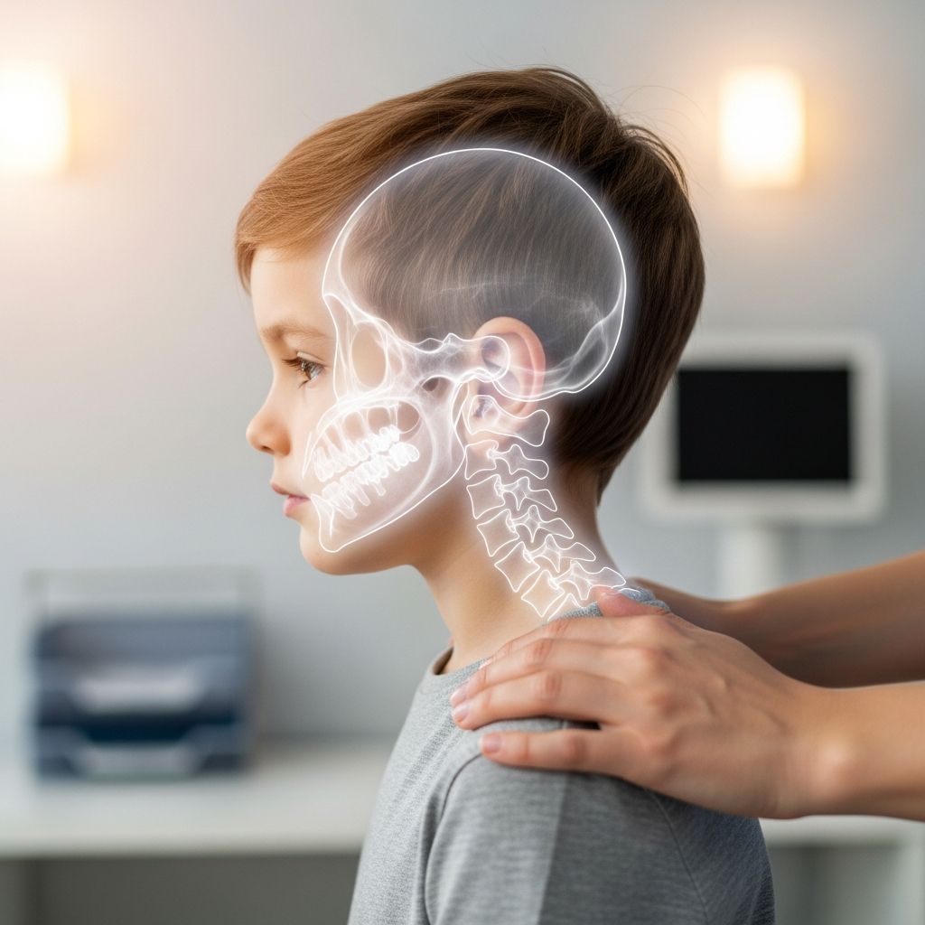
Cervical spine X-rays are a common imaging test used to look at the bones in a child’s neck when there is pain, injury, or concern about how the spine is developing. Understanding why the test is ordered, what happens during the exam, and what the results mean can make the experience much less stressful for you and your child.
What Is the Cervical Spine and Why Does It Matter?
The cervical spine is the part of the backbone that forms the neck. It includes seven stacked bones called vertebrae, labeled C1 through C7, which support the head, protect the spinal cord, and allow the neck to bend and turn.
- C1 (atlas): Helps support the skull.
- C2 (axis): Has a special bony peg (dens) that allows the head to rotate.
- C3–C7: Provide structure, protect nerves, and help with movement.
Inside the vertebrae runs the spinal cord, which carries messages between the brain and the rest of the body. Any serious injury or abnormality in the neck can affect movement, sensation, or even breathing, so doctors are cautious about evaluating this area carefully.
What Is a Cervical Spine X-Ray?
A cervical spine X-ray is a test that uses a small dose of radiation to create images of the bones in the neck. During the exam, an X-ray beam passes through the neck and is captured on a digital detector, producing pictures that show the size, shape, and alignment of the vertebrae.
Cervical spine X-rays are one type of spine X-ray, which can also include images of the thoracic (mid-back) and lumbar (lower back) regions depending on the clinical question.
Why Might a Child Need a Cervical Spine X-Ray?
Doctors may order cervical spine X-rays in children for several reasons. Common situations include:
- Neck injury after trauma
Falls, sports impacts, car crashes, or sudden blows can injure the neck. X-rays help check for fractures, dislocations, or abnormal alignment of the vertebrae.
- Persistent or severe neck pain
When neck pain is ongoing, intense, or associated with stiffness or limited movement, X-rays can help find structural problems like small fractures or early signs of joint disease.
- Abnormal neurologic symptoms
Weakness, numbness, tingling, trouble walking, or changes in coordination may prompt imaging of the cervical spine to see if the spinal cord or nerves are being compressed.
- Suspected congenital or developmental problems
Some children are born with differences in the shape or alignment of the vertebrae. An X-ray can help diagnose these conditions and guide follow-up.
- Monitoring known spine conditions
Children with certain syndromes, skeletal disorders, or previous neck injuries may need periodic X-rays to monitor growth and alignment.
What Can a Cervical Spine X-Ray Show?
This test gives doctors a detailed look at the bony structures in the neck. A cervical spine X-ray can help reveal:
- Fractures (broken bones) in any of the cervical vertebrae.
- Dislocations or subluxations, where vertebrae are out of normal alignment.
- Abnormal curvature or loss of the normal neck curve (lordosis).
- Disc space changes, such as narrowed spaces between vertebrae that may suggest disc problems.
- Changes in bone shape or density related to infection, inflammation, or long-standing conditions.
- Evidence of instability, especially at the junction between C1 and C2 in certain conditions.
Soft tissues (like muscles and ligaments) and the spinal cord itself are not seen in detail on X-ray. If those structures are a concern, doctors may recommend additional tests such as MRI or CT scans.
Types of Cervical Spine X-Ray Views
To fully evaluate the neck, radiology teams usually take more than one view from different angles. Standard views include:
| View | How It’s Taken | What It Shows Best |
|---|---|---|
| Lateral view | Side view of the neck, usually with the child standing, sitting, or lying still. | Overall alignment of the cervical spine from C1 to at least C7, vertebral body height, and prevertebral soft tissue width. |
| AP (anteroposterior) view | Front-to-back image, facing the X-ray tube. | Height and position of vertebrae, spinous processes, and general symmetry. |
| Open-mouth (odontoid) view | Child opens the mouth slightly while the image is taken. | C1 and C2, especially the dens and the spaces around it. |
In some cases, special views—such as flexion/extension lateral images, or a “swimmer’s view” to see the lower neck when shoulders cover it—may be used depending on how well the standard views show the area of concern.
How Safe Are Cervical Spine X-Rays for Children?
X-rays use ionizing radiation, so doctors carefully weigh the benefits and risks before ordering them for children. Modern equipment and strict safety standards mean that the radiation dose from a single cervical spine X-ray study is typically low.
Key safety points include:
- Justification: The test is ordered only when it is likely to provide important information for diagnosis or treatment.
- Lowest reasonable dose: Radiology teams use pediatric settings, limiting exposure to the smallest amount needed for a clear image (the “ALARA” principle—As Low As Reasonably Achievable).
- Shielding and positioning: Lead aprons or thyroid shields may be used when appropriate, and only the body area being examined is exposed.
- Limiting repeat images: Careful positioning and clear instructions help avoid the need to repeat X-rays.
When neck injury or disease is suspected, the information gained from X-rays usually far outweighs the small potential risk from radiation exposure.
How to Prepare Your Child
Most children do not need any special medical preparation before a cervical spine X-ray, but emotional preparation makes a big difference. Helpful steps include:
- Explain in simple terms
Describe the test as a “special picture of your neck bones” that doesn’t hurt. Let your child know that they will need to hold still for a few seconds at a time.
- Choose comfortable clothing
Have your child wear soft, loose clothes without metal zippers, snaps, or necklaces. Metal can block X-rays and may need to be removed.
- Bring comfort items
A favorite small toy, blanket, or stuffed animal can help your child feel more secure while waiting and during the test.
- Answer questions honestly
If your child asks whether it will hurt, you can say that the machine does not hurt, but staying still may feel a bit uncomfortable for a short time.
What Happens During the Exam?
Cervical spine X-rays are usually performed in a radiology or imaging department. The entire visit often takes less than 30 minutes, with the actual exposure time lasting just a fraction of a second for each picture.
Step-by-step overview
- Check-in
You will be asked about your child’s medical history, recent injuries, and any possibility of pregnancy in teen patients.
- Positioning
A radiologic technologist helps your child stand, sit, or lie in the correct position for each view. They may gently adjust the head and shoulders to get a clear picture.
- Use of shields
When appropriate, a lead apron or shield is placed over parts of the body not being imaged to limit exposure.
- Staying still
Your child will be asked to hold very still and sometimes to hold their breath briefly. Even small movements can blur the image.
- Taking the images
The technologist steps behind a protective barrier and activates the X-ray. Your child will not feel the X-rays as they pass through.
- Checking image quality
The technologist checks that all necessary structures are visible. If something is unclear, an additional image may be taken.
Parents are often allowed to stay in the room, wearing a protective apron, to comfort younger children, as long as they are not pregnant.
After the X-Ray: Results and Next Steps
Once the images are taken, a specially trained doctor called a radiologist reviews them, looking systematically at the bones, spaces between them, and overall alignment.
Your child’s doctor may receive a report within hours to a day, depending on urgency. In emergency situations, the images are reviewed immediately.
Possible outcomes
- Normal study
No fractures, dislocations, or concerning abnormalities are seen. Your child’s symptoms may be due to muscle strain, minor soft-tissue injury, or another cause not visible on X-ray.
- Abnormal findings
The report may describe a fracture, misalignment, abnormal curve, or unusual bone appearance. Your child’s doctor will explain what this means and discuss treatment or further testing.
- Unclear or incomplete
Sometimes, especially if the child moved or the shoulders block part of the lower neck, it can be difficult to see all vertebrae clearly. In that case, additional X-ray views or another imaging test such as CT or MRI may be recommended.
When Is CT or MRI Used Instead of X-Ray?
X-rays are usually the first step for many neck problems because they are quick, widely available, and use relatively low doses of radiation. However, other imaging methods may be better in certain situations:
- Computed tomography (CT)
CT scans provide much more detailed cross-sectional images of bone and can better show complex fractures or subtle alignment issues. CT uses higher radiation doses than plain X-rays, so doctors reserve it for cases where detailed information is essential, such as serious trauma.
- Magnetic resonance imaging (MRI)
MRI does not use ionizing radiation. Instead, it uses a strong magnet and radio waves to create detailed images of soft tissues, including the spinal cord, discs, ligaments, and nerves. MRI may be ordered if there are neurologic symptoms or concern for spinal cord injury that X-rays and CT cannot fully explain.
Helping Your Child Cope With Anxiety
Even though cervical spine X-rays are painless, the unfamiliar environment and need to remain still can be stressful. Strategies that often help include:
- Practice staying still: Turn it into a game at home—see who can freeze like a statue for 10–15 seconds.
- Use age-appropriate explanations: For younger kids, compare the machine to a “big camera”; for older children, you can describe how the pictures help doctors see the bones.
- Offer choices when possible: Let your child choose which stuffed animal to bring or whether you hold their hand during the test.
- Stay calm yourself: Children often mirror a parent’s mood. Speaking softly and confidently can reassure them.
Frequently Asked Questions (FAQs)
Will my child feel anything during a cervical spine X-ray?
No. Your child will not feel the X-rays. The only discomfort is usually from holding still or keeping the neck in one position for a short time.
How long does the test take?
The actual X-ray exposures take less than a second each. With positioning and preparation, most appointments are finished within about 15–30 minutes.
Is sedation ever needed?
Sedation is rarely used for simple X-rays because the test is quick and noninvasive. In very young or extremely anxious children, staff may use extra support, distraction, or immobilization devices to help the child hold still instead of medication.
How much radiation will my child receive?
The radiation dose from a cervical spine X-ray is generally low and is kept as minimal as possible using pediatric protocols. The radiology team balances any small risk from radiation against the need to diagnose a potentially serious neck problem.
Can I stay with my child during the exam?
In most facilities, a parent or caregiver can remain in the room, wearing a lead apron for protection, unless they are pregnant. Ask the staff about their policy when you arrive.
What if the X-ray is normal but my child still hurts?
A normal X-ray does not mean your child’s pain is not real. Many causes of neck pain—such as muscle strain, ligament sprain, or minor disc irritation—do not show up on X-ray. Your child’s doctor will consider the X-ray results along with the physical exam and symptoms to decide on next steps.
How quickly will we get the results?
In urgent cases, such as after an accident, a doctor may give you a preliminary interpretation almost immediately. In less urgent situations, the formal radiology report is often available within a day, and your child’s doctor will review the findings with you.
References
- Spine X-Ray: What It Is, Purpose, Procedure, Results & Types — Cleveland Clinic. 2022-08-05. https://my.clevelandclinic.org/health/diagnostics/10229-spine-x-ray
- Cervical Spine X-ray Interpretation — Geeky Medics. 2023-03-15. https://geekymedics.com/cervical-spine-x-ray-interpretation-osce-guide/
- Radiographic Positioning Techniques for the Cervical Spine — AuntMinnie / Radiology Practice Management. 2019-06-10. https://www.auntminnie.com/practice-management/radiologic-technologist/article/15564550/radiographic-positioning-techniques-for-the-cervical-spine
- How to Read a C-Spine X-ray — NewYork-Presbyterian Hospital. 2021-01-12. https://www.nyp.org/professionals/emergency-medicine/how-to-read-emergency-images/how-to-read-a-c-spine-film
- How to Read C-Spine X-Ray — International Emergency Medicine Education Project. 2018-04-02. https://iem-student.org/how-to-read-c-spine-x-ray/