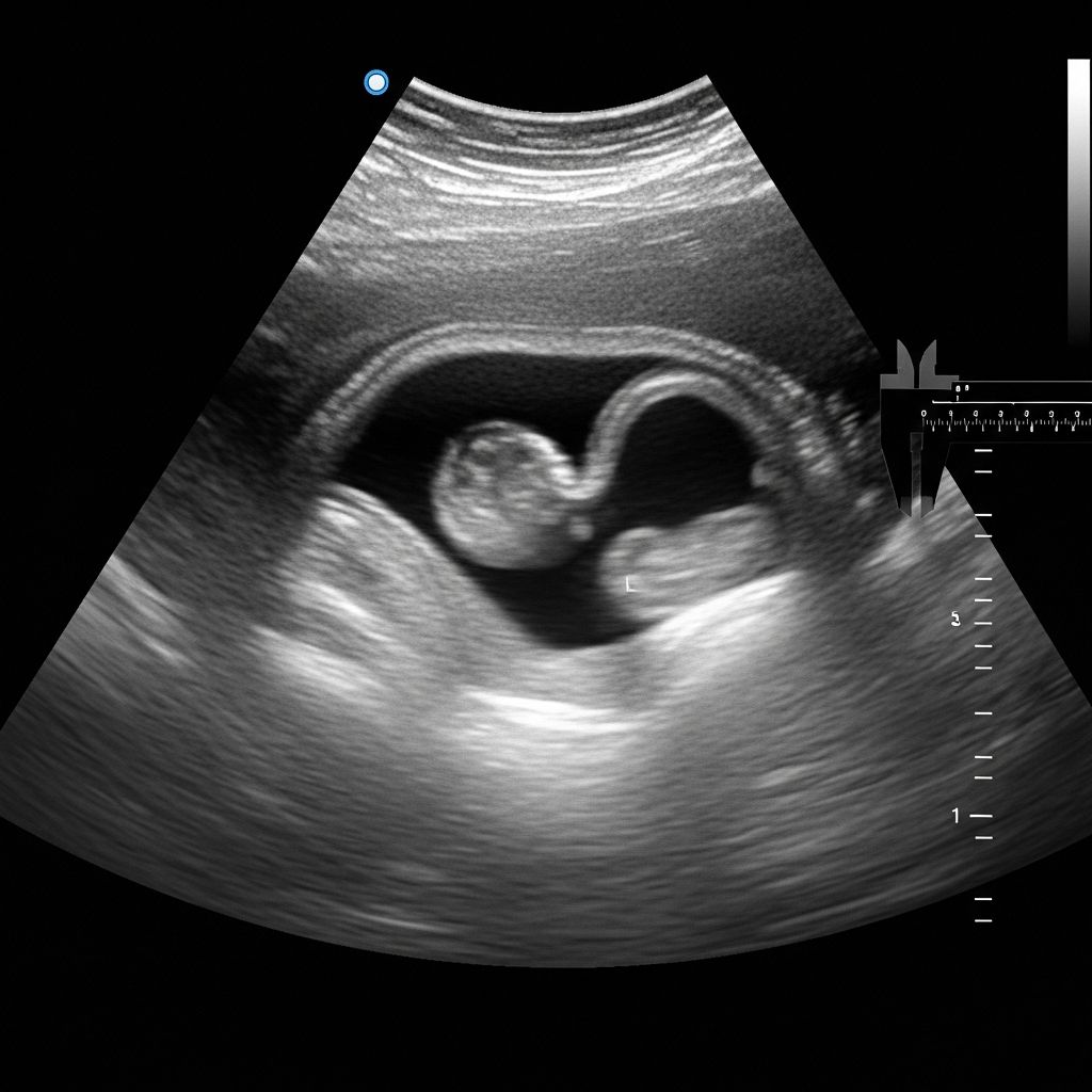
Navigating Early Pregnancy Ultrasound Concerns
One of the most emotionally charged moments in early pregnancy occurs when an ultrasound fails to detect a fetal heartbeat. For expectant parents, this discovery can trigger immediate anxiety and worst-case assumptions. However, the absence of a detectable heartbeat at this early stage does not necessarily indicate a miscarriage or other serious complications. Understanding the various factors that influence heartbeat detection can help parents approach this situation with more clarity and informed perspective.
The detection of a fetal heartbeat represents one of the earliest confirmations of a viable pregnancy. Most healthcare providers and expectant parents eagerly await this milestone during the first prenatal ultrasound. Yet in approximately 15 to 20 percent of early pregnancy ultrasounds, no heartbeat is initially detected. While this statistic can feel alarming, research and clinical experience demonstrate that many of these pregnancies progress normally and result in healthy births.
The Timeline of Fetal Heartbeat Detection
Understanding when a fetal heartbeat should be detectable helps contextualize why early ultrasounds might not reveal one. The timeline varies somewhat based on the measurement method and ultrasound technology used.
Fetal cardiac activity typically becomes detectable between 41 and 43 days of gestation, with fetal heart rates ranging from 96 to 120 beats per minute. By the eighth week of pregnancy, fetal heart activity can be detected through abdominal ultrasound with high certainty. However, transvaginal ultrasound, which inserts the probe into the vagina rather than across the abdomen, can identify cardiac activity earlier and with greater sensitivity than transabdominal ultrasound.
Doppler signals, which detect blood flow from the fetal heart and associated blood vessels, can typically identify cardiac activity after six weeks of pregnancy. From the twentieth week onward, a trained healthcare provider can often detect the fetal heartbeat using a standard stethoscope during physical examination. A progressively stronger heartbeat as pregnancy advances generally indicates healthy fetal development and appropriate growth.
Technical and Measurement Factors Affecting Detection
Several technical and clinical factors can influence whether a heartbeat appears on an early pregnancy ultrasound, independent of fetal viability.
Gestational Age Calculation Errors
One of the most common reasons for absent heartbeat detection involves inaccurate calculation of gestational age. Pregnancy dating typically begins from the first day of the last menstrual period, but this method has inherent limitations. If ovulation occurred later than average, or if menstrual cycles are irregular, the actual age of the embryo may be considerably younger than dates suggest.
Late implantation of the fertilized egg also affects developmental timeline. When implantation occurs later than typical, the embryo appears developmentally younger than expected based on last menstrual period dating. In such cases, waiting one to two weeks and repeating the ultrasound often reveals a normally developing embryo with a clearly visible heartbeat. Many cases of “no heartbeat” at six to seven weeks actually represent pregnancies that are only at the four to five week developmental stage.
Ultrasound Equipment and Operator Expertise
The quality of ultrasound equipment significantly impacts heartbeat detection capability. Older or lower-resolution machines may struggle to visualize a tiny embryonic heart. The expertise and experience of the ultrasound technician also plays a crucial role. A skilled sonographer with access to modern equipment and transvaginal imaging capability is far more likely to detect a heartbeat than a less experienced operator using only transabdominal imaging.
Fetal Positioning and Maternal Factors
The position of the embryo within the uterus can affect visualization. If the embryo positions itself in a way that makes the heartbeat difficult for ultrasound waves to detect, a heartbeat may appear absent even though the fetus is developing normally. Similarly, maternal factors such as a tilted or retroverted uterus, elevated body mass index, or uterine fibroids can create imaging challenges that impede heartbeat detection on transabdominal ultrasound.
Developmental Variations Among Pregnancies
Embryonic development does not follow a perfectly uniform timeline across all pregnancies. Individual variations in fetal development occur naturally and commonly.
Some embryos develop a detectable heartbeat slightly later than the statistical average without any pathological cause. These developmental variations may reflect genetic factors, individual metabolic differences, or other normal biological variation. Research indicates that some fetuses demonstrate slower early development yet continue progressing normally throughout pregnancy, eventually achieving all developmental milestones and resulting in full-term, healthy births.
In approximately 10 to 15 percent of cases where no heartbeat is detected at six weeks, a heartbeat becomes clearly visible one week later. This pattern strongly suggests that the initial absence reflected developmental timing rather than pregnancy loss. Parents in this situation benefit enormously from understanding that delayed heartbeat detection does not doom the pregnancy.
When Absent Heartbeat Signals Genuine Concern
While many cases of absent heartbeat resolve favorably with follow-up ultrasound, some situations warrant more serious consideration of miscarriage or other pregnancy complications.
The Role of Human Chorionic Gonadotropin Levels
When no heartbeat is detected, healthcare providers may order blood tests measuring human chorionic gonadotropin (HCG). This hormone rises predictably during early pregnancy. Normal, appropriately rising HCG levels in the context of no detected heartbeat suggest the pregnancy may still be viable, and the absence likely reflects timing issues rather than fetal demise. Declining HCG levels, conversely, indicate that the pregnancy is likely not viable and miscarriage may be occurring or imminent.
The Blighted Ovum Diagnosis
A blighted ovum, also called an anembryonic pregnancy, occurs when a gestational sac develops normally but no embryo forms within it. In these cases, repeated ultrasounds one to two weeks apart will continue showing only an empty sac with no fetal development and no heartbeat. Once this pattern becomes clear, no supportive treatment can restore the pregnancy, as it is fundamentally non-viable from the beginning.
Missed Miscarriage Characteristics
A missed miscarriage involves fetal death but retention of the pregnancy tissue within the uterus. Ultrasound typically shows a pregnancy sac and fetal tissue, but without a visible heartbeat. These pregnancies require medical management to safely remove tissue and prevent infection or other complications.
Recommended Next Steps and Follow-Up Care
The standard approach when no heartbeat is detected involves several sequential steps designed to obtain clarity without rushing to conclusions.
Initial Response and Waiting Period
If no heartbeat appears at six to seven weeks of gestation and maternal symptoms are normal, most healthcare providers recommend waiting one to two weeks before repeating the ultrasound. This waiting period allows for potential advancement in fetal development and provides clearer information about whether the pregnancy is progressing. During this time, HCG blood tests may be ordered to confirm that hormone levels are rising appropriately.
Repeat Ultrasound Protocol
Follow-up ultrasound typically employs transvaginal imaging, which provides superior resolution compared to transabdominal approaches, particularly in very early pregnancy. The repeat study should be performed at least one week after the initial ultrasound to allow sufficient development time to become apparent if the pregnancy is viable.
When Urgent Investigation Becomes Necessary
Certain maternal symptoms warrant immediate rather than delayed follow-up. If a mother experiences severe lower abdominal pain, heavy vaginal bleeding, or sudden cessation of pregnancy symptoms such as morning sickness, medical evaluation should occur promptly rather than waiting for a scheduled repeat ultrasound. These symptoms may indicate miscarriage in progress or other complications requiring immediate intervention.
Testing After Twelve Weeks
If ultrasound at twelve weeks or beyond still shows no fetal heartbeat, healthcare providers will pursue more urgent investigation. At this gestational age, the absence of a heartbeat more strongly suggests either a non-viable pregnancy or significant fetal abnormalities. Urgent HCG testing and potentially additional imaging will be ordered to confirm the situation and discuss appropriate management options.
Genetic and Medical Considerations
In approximately 90 percent of cases where fetal heartbeat is permanently absent despite appropriate gestational age and development, genetic abnormalities account for the non-viability. Chromosomal problems such as trisomy, monosomy, or other genetic errors occur randomly during embryo formation and cannot be prevented through any parental action or lifestyle choice.
When a miscarriage occurs due to genetic factors, healthcare providers may recommend genetic testing of the miscarried tissue to identify the specific abnormality. This information, while not changing the current outcome, provides valuable understanding for future pregnancy planning. Additionally, some couples benefit from blood testing to evaluate their own chromosomes and assess whether parental factors contributed to the loss.
Emotional Support and Coping Resources
The experience of learning that no heartbeat is detected creates significant emotional turmoil. Parents should understand that their anxiety and distress are completely valid responses to this frightening situation. The uncertainty of not knowing whether the pregnancy will continue creates substantial psychological burden.
Families benefit from support from healthcare providers who communicate compassionately about the situation, explain the rationale for follow-up timing, and avoid prematurely suggesting miscarriage when the actual situation remains uncertain. Support from friends, family members, and sometimes professional counselors or support groups helps parents navigate this challenging period. Organizations dedicated to miscarriage and pregnancy loss provide evidence-based information and emotional support resources.
Frequently Asked Questions
Does no heartbeat at six weeks always mean miscarriage?
No. In 10 to 15 percent of cases where no heartbeat is detected at six weeks, a heartbeat becomes visible one week later. Many cases reflect incorrect dating rather than miscarriage. Only when the pattern persists across multiple ultrasounds or HCG levels decline does miscarriage become likely.
Should I request an ultrasound as soon as possible if worried?
Very early repeated ultrasounds may not provide additional clarity and can increase anxiety. Healthcare providers typically recommend waiting one to two weeks to allow fetal development to progress. This approach yields more definitive information than rushing to repeat imaging.
What does a normal HCG level mean when heartbeat is absent?
Normal and appropriately rising HCG suggests the pregnancy is likely still viable. It indicates hormonal support for pregnancy is adequate, and the absent heartbeat probably reflects developmental timing rather than fetal death.
Can maternal stress or lifestyle choices cause absent heartbeat?
No. Absent heartbeat in early pregnancy reflects either technical factors preventing detection or fetal viability issues caused by genetic or developmental factors. Parental stress, exercise, diet, or other lifestyle choices do not cause these outcomes.
At what point should I accept that miscarriage has occurred?
When ultrasound at twelve weeks or beyond continues showing no heartbeat, when serial HCG levels decline, or when your healthcare provider confirms that the pregnancy sac remains empty and unchanging across multiple ultrasounds, the diagnosis of non-viable pregnancy becomes definitive.
Are there treatments to restore a heartbeat once it appears absent?
No medical treatment can restore fetal cardiac activity if the fetus is not viable. However, in many cases, waiting and repeat ultrasound reveal that the heartbeat was never absent—it simply wasn’t detected initially due to technical factors.
Moving Forward with Confidence
The absence of a fetal heartbeat on an early pregnancy ultrasound creates one of the most anxiety-provoking experiences in obstetrics. However, understanding that this finding does not automatically indicate miscarriage, and that many such pregnancies progress normally, can help parents navigate this uncertainty with greater emotional resilience. Close follow-up with a trusted healthcare provider, attention to concerning symptoms, and realistic expectations about ultrasound limitations all contribute to appropriate management and optimal outcomes for these pregnancies.
References
- Correct Understanding of Ultrasound Results Without Fetal Heart — Vinmec International Hospital. 2024. https://www.vinmec.com/eng/blog/correct-understanding-of-ultrasound-results-without-fetal-heart-en
- No Heartbeat at 6 Weeks — International Ultrasound Services London. 2024. https://www.iuslondon.co.uk/blog/pregnancy/no-heartbeat-at-6-weeks/
- Is No Fetal Heartbeat at 6 Weeks of Pregnancy a Sign of Miscarriage? — Nishanti IVF Care. 2024. https://www.nishantivfcare.com/is-no-fetal-heartbeat-at-6-weeks-of-pregnancy-a-sign-of-miscarriage/
- How Late Can Heartbeat Start in Pregnancy — The Ova Care. 2024. https://theovacare.com/how-late-can-heartbeat-start-in-pregnancy/
- Missed Miscarriage — Miscarriage Association. 2024. https://www.miscarriageassociation.org.uk/information/miscarriage/missed-miscarriage/
- Patience is Key: Understanding the Timing of Early Ultrasounds — UT Southwestern Medical Center. 2024. https://utswmed.org/medblog/patience-key-understanding-timing-early-ultrasounds/