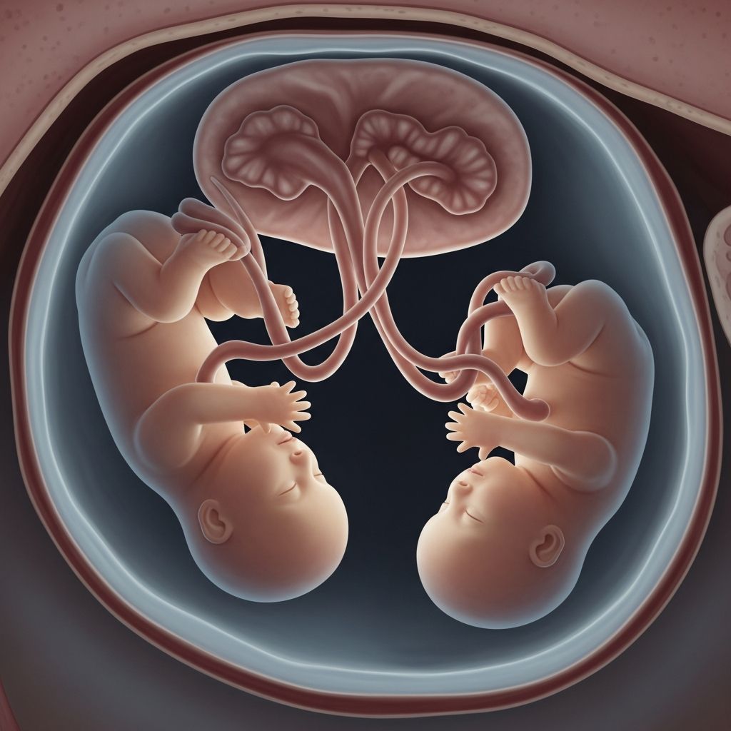
Monoamniotic twins, also known as mono-mono twins, represent the rarest form of identical twin pregnancy, occurring in approximately 1 in 10,000 pregnancies or about 1% of monozygotic twins. These twins share a single amniotic sac and placenta, creating unique and severe risks that demand specialized prenatal care.
Understanding the Biology of Monoamniotic Twins
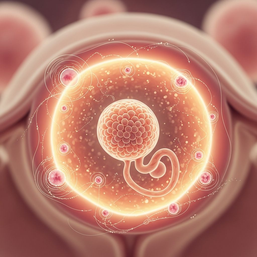
Identical twins form when a single fertilized egg splits into two embryos. The timing of this split determines the placental and amniotic sac arrangement. If the split happens more than 13 days after fertilization, the embryos remain within one chorion (outer membrane) and one amnion (inner sac), resulting in a monochorionic-monoamniotic (MCMA) pregnancy.
This configuration means both babies develop in the same fluid-filled space without any dividing membrane. They share one placenta, with umbilical cords originating close together—often just inches apart—which heightens the chance of complications.
- Key biological features: Single placenta (monochorionic), single amniotic sac (monoamniotic).
- Prevalence: Less than 1% of all twin pregnancies in the U.S.
- Identical nature: Always monozygotic, sharing nearly identical DNA.
Primary Dangers in Monoamniotic Pregnancies
The shared environment exposes monoamniotic twins to dangers beyond those of typical twin pregnancies. The absence of a membrane allows free movement, but it also facilitates life-threatening issues.
Cord Entanglement and Compression
The most critical risk is umbilical cord entanglement. With cords emerging from the same placental site and traversing the same sac, they can twist, knot, or compress, interrupting blood and oxygen flow to one or both fetuses. This can occur suddenly without warning, leading to fetal distress or demise.
- Entanglement risk peaks after 24-28 weeks when twins become more active.
- Compression mimics a kinked hose, starving babies of nutrients.
- Historical mortality from this alone approached 50%, though modern monitoring has improved outcomes.
Twin-to-Twin Transfusion Syndrome (TTTS)
In monochorionic pregnancies, placental blood vessel connections can cause unequal blood sharing. TTTS affects 10-15% of such cases, where one twin (donor) pumps blood to the other (recipient) via arteriovenous anastomoses. The donor becomes anemic and growth-restricted, while the recipient risks heart failure and fluid overload.
Advanced TTTS stages lead to oligohydramnios (low fluid) in the donor sac and polyhydramnios (excess fluid) in the recipient, further compressing cords.
Other Complications
| Complication | Description | Risk Level |
|---|---|---|
| Selective Fetal Growth Restriction (sFGR) | One twin receives less placental share, stunting growth. | High |
| Preterm Labor | 50% chance before 37 weeks, often earlier. | Very High |
| Congenital Anomalies | Increased structural defects due to late splitting. | Elevated |
| Maternal Risks | Preeclampsia, anemia, hemorrhage. | Moderate-High |
Perinatal mortality for monoamniotic twins reaches 8-20% or higher without intervention, far exceeding dichorionic twins.
Diagnosing Monoamniotic Twins Early
Routine ultrasound around 10-14 weeks often reveals monoamniotic twins. Key signs include a single placental mass, absent dividing membrane, and intertwined cords.
- First trimester: Nuchal translucency scan may show shared sac.
- Second trimester: Detailed anatomy scan confirms no membrane and cord proximity.
- Fetal echocardiography: Screens for TTTS-related heart issues.
Early diagnosis shifts care to maternal-fetal medicine specialists at high-risk centers equipped with NICU facilities.
Intensive Monitoring Protocols
Once identified, monoamniotic pregnancies require hospitalization from 24-26 weeks or intensive outpatient surveillance. The goal: balance prematurity risks against sudden cord events.
- Inpatient monitoring: Twice-daily non-stress tests (NST) track heart rates; biophysical profiles assess movements, tone, breathing, and fluid.
- Ultrasound frequency: Weekly for growth, Doppler for blood flow, amniotic fluid index.
- Maternal surveillance: Blood pressure checks for preeclampsia; steroids at 24-28 weeks for lung maturity.
Any heart rate deceleration prompts emergency evaluation. Bed rest minimizes twin activity that could worsen cord issues.
Timing and Methods of Delivery
Vaginal delivery is rare due to risks; cesarean section is standard. Debate centers on timing: expectant management vs. planned early delivery.
- Conservative approach: Monitor until 32-34 weeks if stable, minimizing prematurity.
- Early delivery: At 32 weeks to preempt cord accidents, supported by recent studies showing reduced fetal loss.
- Emergency C-section: Indicated for distress, as in cases where knots form suddenly.
Post-32 weeks, the risk of respiratory distress from prematurity drops, while cord risks rise exponentially.
Neonatal Care for Preterm Monoamniotic Twins
Delivered often before 34 weeks, these babies require Level III/IV NICU care. Common needs include respiratory support, feeding assistance, and infection monitoring.
- Short-term: Surfactant therapy, CPAP/ventilation for lungs; IV nutrition.
- Long-term: Follow-up for neurodevelopment, as preterm mono-mono twins face higher cerebral palsy risk.
Survival exceeds 80-90% in expert centers today, a vast improvement from historical rates.
Real-Life Outcomes and Survivor Stories
Cases like Kate and Annie Carlson highlight success: born at 30 weeks via emergency C-section after cord knot detection, they thrived in NICU. Similarly, specialized teams at hospitals like Samitivej report positive outcomes with vigilant care.
Challenges persist; some pregnancies end in loss despite monitoring, underscoring the gravity.
Emotional and Practical Support for Parents
Facing a monoamniotic diagnosis brings anxiety. Support includes:
- Counseling from perinatologists and psychologists.
- Connections to twin parent groups.
- Financial planning for extended hospitalization.
Preparation involves hospital tours, birth plans focused on C-section, and lactation support for NICU feeding.
Frequently Asked Questions (FAQs)
What causes monoamniotic twins?
A late split of the inner cell mass after day 13 post-fertilization traps both embryos in one sac.
Can monoamniotic twins be delivered vaginally?
Rarely; C-section is preferred to control timing and avoid cord prolapse.
What is the survival rate today?
Over 80% in high-risk centers with modern monitoring, down from 50% mortality historically.
Is bed rest always required?
Often from 24 weeks inpatient to detect issues promptly.
Can TTTS be treated in mono-mono twins?
Fetoscopic laser surgery is challenging due to single sac but possible in select cases.
Advances in Care and Future Outlook
Ongoing research explores optimal delivery timing via trials like planned 32-week C-sections vs. monitoring. Fetal surgery innovations and better imaging promise further gains. For now, referral to tertiary centers is crucial.
(Word count: 1678)
References
- Monochorionic-Monoamniotic (MCMA) Twins — Samitivej Hospitals. 2023. https://www.samitivejhospitals.com/article/detail/monochorionic-monoamniotic-mcma-twins
- Planned early delivery versus expectant management for monoamniotic twin pregnancies — PMC / Cochrane Review. 2022-04-12. https://pmc.ncbi.nlm.nih.gov/articles/PMC8947902/
- Monoamniotic Twins: How Does The Condition Affect Your Pregnancy? — WebMD. 2023. https://www.webmd.com/baby/what-are-monoamniotic-twins
- Twins who shared amniotic sac, a potentially deadly condition, now home with mom and dad — Stanford Medicine. 2013-01-28. https://med.stanford.edu/news/all-news/2013/01/twins-who-shared-amniotic-sac-a-potentially-deadly-condition-now-home-with-mom-and-dad.html
- Mono mono twins — Hamilton Health Sciences. 2023. https://www.hamiltonhealthsciences.ca/share/mono-mono-twins/
- Birth Defects In Monochorionic Twins — Children’s Wisconsin. 2023. https://childrenswi.org/find-care/fetal-concerns/birth-defects-in-monochorionic-twins
- Twin Pregnancy Complications — UCSF Fetal Treatment Center. 2023. https://fetus.ucsf.edu/twin-pregnancy-complications/