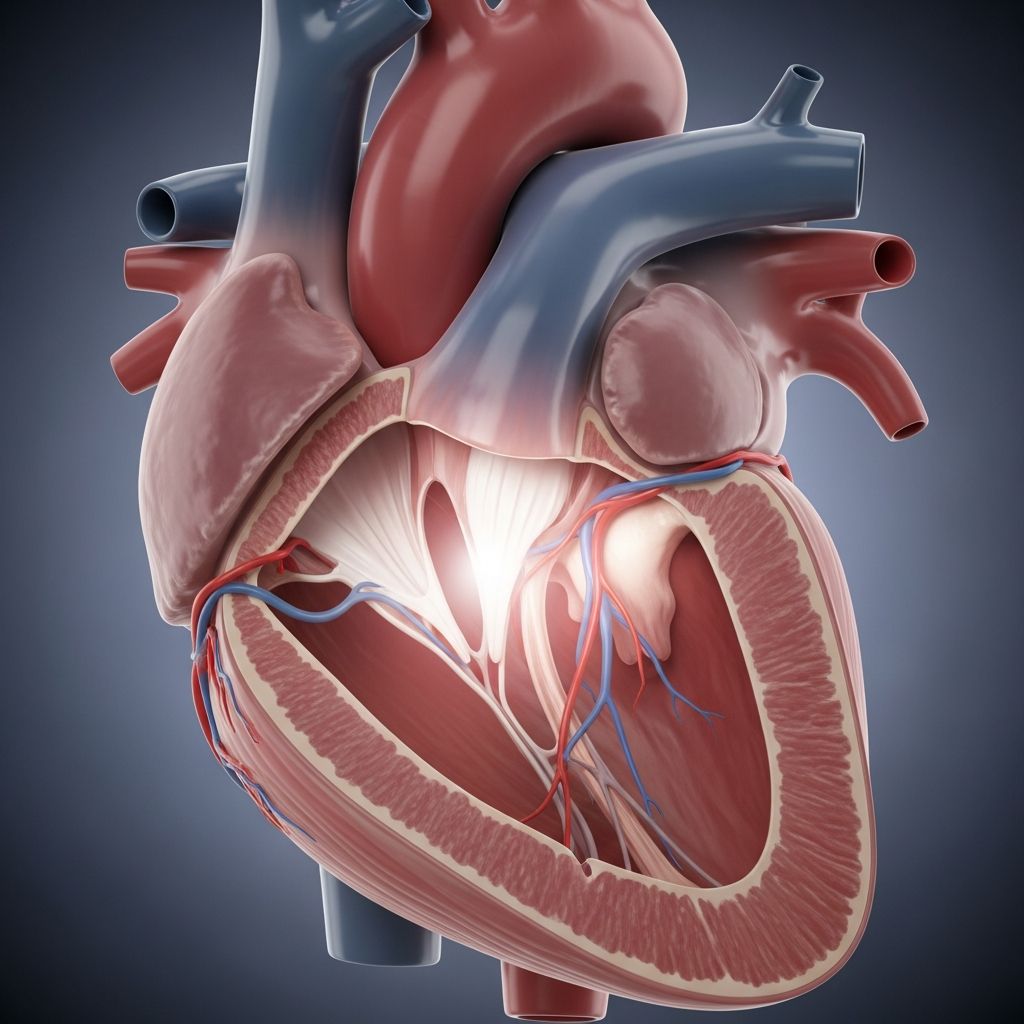
The Left Atrium: A Vital Chamber in Your Heart
Your heart is a remarkable organ composed of four distinct chambers that work in perfect harmony to pump blood throughout your entire body. Among these chambers, the left atrium plays a critical role in ensuring that oxygen-rich blood reaches every cell and tissue. Understanding how this chamber functions helps you appreciate the complexity of your cardiovascular system and why maintaining heart health is essential for overall wellness.
The left atrium is one of two upper chambers of the heart, positioned on the left side and slightly toward the back of the chest cavity. While it may seem small compared to the more muscular ventricles below it, this chamber performs vital functions that are absolutely necessary for life. Its primary responsibilities include receiving oxygenated blood from the lungs and delivering that blood to the left ventricle, which then pumps it throughout the body.
Anatomical Position and Structure
The left atrium occupies a specific location within the heart’s architecture. Positioned behind and slightly to the left of the right atrium, it sits above the left ventricle and forms part of the heart’s posterior surface. In anatomical terms, the left atrium contributes to what is called the anatomical base of the heart, which points toward the back of the chest cavity.
The walls of the left atrium are notably thicker than those of the right atrium. This structural difference exists because the left atrium must handle higher pressures as it receives and pumps oxygenated blood, requiring more muscular tissue to perform this demanding task. The myocardial wall—the muscular layer that contracts to move blood—is constructed with three distinct regions: the venous portion that receives blood from the pulmonary veins, the auricular region which forms the left atrial appendage, and the vestibular section.
A defining feature of the left atrium is the presence of pectinate muscles, which are small muscle bundles visible as ridges on the interior surface. These muscles are found primarily in the auricular portion of the left atrium and contribute to its contractile strength. The smooth venous region, by contrast, has a streamlined appearance that facilitates efficient blood flow.
The Four Pulmonary Veins: Pathways of Oxygenated Blood
The left atrium receives oxygenated blood through a specialized network of vessels called the pulmonary veins. Unlike most veins in the body that carry deoxygenated blood, the pulmonary veins are unique in that they transport oxygen-rich blood from the lungs back to the heart. Typically, four pulmonary veins connect to the left atrium—two from the right lung and two from the left lung—though anatomical variations do occur in some individuals where these veins may merge into a common trunk before entering the chamber.
These veins open into the left atrium through small openings called ostia, which are strategically located in the upper, back, and outer portion of the atrial wall. The positioning of these openings ensures optimal blood flow dynamics and allows the chamber to efficiently collect the oxygen-rich blood returning from the lungs. The smooth muscular surface surrounding these openings helps facilitate smooth blood entry into the chamber.
The Mitral Valve: Gateway to the Left Ventricle
Once blood has accumulated in the left atrium, it must be transported to the left ventricle for further circulation throughout the body. This transfer occurs through a specialized one-way valve called the mitral valve, also known as the left atrioventricular valve. This valve functions as a biological gate, permitting blood to flow only in one direction—from the atrium into the ventricle—and preventing backflow.
The mitral valve consists of two leaflets or cusps that open during atrial contraction and close when the left ventricle begins to contract. This coordinated movement ensures that blood moves progressively through the heart chambers without returning to previous chambers. A properly functioning mitral valve is essential for efficient circulation, and problems with this valve can significantly impact heart function and overall health.
The Left Atrial Appendage: A Specialized Pouch
An intriguing structural feature of the left atrium is a small, ear-shaped muscular pouch located in the upper portion called the left atrial appendage (LAA), also referred to as the left auricle. This structure has a complex internal architecture with a trabeculated design, meaning it contains small ridges and crevices. Interestingly, the shape of the left atrial appendage varies among individuals, and medical professionals classify these variations into four main morphological types based on appearance.
The left atrial appendage serves multiple important functions. It acts as a decompression chamber during certain phases of the heartbeat, particularly when pressure within the left atrium rises. Additionally, this structure is involved in regulating body fluid balance by secreting hormones called natriuretic peptides, specifically atrial natriuretic peptide and brain natriuretic peptide. These hormones enter the bloodstream and help regulate blood pressure and fluid balance throughout the body.
The Electrical Conduction System and the Left Atrium
While the left atrium’s primary function is mechanical—receiving and pumping blood—it also plays an important role in the heart’s electrical system. The electrical signal that coordinates heart contractions originates in the right atrium at a specialized structure called the sinoatrial node. This signal travels through the right atrial muscle and reaches the atrioventricular node, which serves as a relay station for electrical impulses.
The electrical signal then travels to the left atrium through a structure called Bachmann’s bundle, a specialized pathway of conductive tissue that bridges the two atria. This ensures that both atria contract in a coordinated fashion, allowing them to work together to fill the ventricles efficiently. The timing of this electrical activity is crucial—a slight delay in the atrioventricular node gives the atria time to fully empty before the ventricles contract, optimizing blood flow through the heart.
The Interatrial Septum: Separating Right from Left
The left atrium is separated from the right atrium by a wall of tissue called the interatrial septum. This muscular partition prevents mixing of oxygen-poor blood in the right atrium with oxygen-rich blood in the left atrium. Within this septum is a depression called the fossa ovalis, which is a remnant of fetal circulation. Before birth, the heart functions differently because the lungs are not yet inflated with air, so a small opening (foramen ovale) allows blood to bypass the lungs by flowing directly from the right to the left atrium. After birth, this opening typically closes, though in some individuals, a small probe-patent foramen ovale may persist without causing problems.
The Contraction Cycle: How the Left Atrium Pumps Blood
The left atrium functions through a carefully orchestrated contraction cycle that coordinates with the rest of the heart. When blood from the pulmonary veins fills the left atrium, the chamber expands. In response to the electrical signal from Bachmann’s bundle, the muscular walls of the left atrium contract, forcefully pushing blood through the mitral valve into the left ventricle below.
This contraction is essential because it contributes to filling the left ventricle and preparing it for its powerful contraction. The left ventricle then contracts and pumps oxygenated blood into the aorta, the large artery that distributes blood to the entire body. After the left ventricle contracts, the atria relax and begin filling with blood again, starting the cycle anew with each heartbeat.
Blood Supply to the Left Atrium
Like all living tissue, the left atrium requires its own blood supply to function properly. The coronary arteries—specialized blood vessels that supply the heart muscle itself—provide nutrients and oxygen to the atrial tissue. The left main coronary artery and its branches deliver approximately 80% of the blood supply to the heart, ensuring that the left atrium receives adequate oxygenation and nutrients to maintain its function.
Common Conditions Affecting the Left Atrium
Several medical conditions can affect the left atrium and impact heart function. One common condition is mitral valve prolapse, in which the mitral valve does not close properly, allowing some blood to leak backward into the left atrium during ventricular contraction. Another significant condition is atrial fibrillation, an irregular heart rhythm in which the left atrium fibrillates or quivers instead of contracting effectively. This irregular motion can lead to blood stasis (slowness of blood flow) within the chamber and increase the risk of blood clot formation.
The left atrial appendage becomes particularly important in atrial fibrillation because this is where blood clots most commonly form when the atrium is not contracting properly. In some cases, surgical procedures may be performed to occlude or close the left atrial appendage to reduce stroke risk in patients with atrial fibrillation.
Why Understanding the Left Atrium Matters
Learning about the left atrium provides insight into how your body maintains circulation and delivers life-sustaining oxygen to every cell. This knowledge becomes especially valuable when healthcare providers discuss heart conditions or when you encounter information about cardiac health. Understanding the normal structure and function of the left atrium helps you recognize when something might be amiss and appreciate the importance of maintaining cardiovascular health through exercise, proper nutrition, and regular medical checkups.
Key Points to Remember
- The left atrium is one of four heart chambers, located on the left posterior side of the heart
- It receives oxygenated blood from the lungs via four pulmonary veins
- The mitral valve controls blood flow from the left atrium to the left ventricle
- The left atrial appendage is a small pouch involved in decompression and hormone secretion
- Thicker walls in the left atrium reflect higher pressures compared to the right atrium
- The left atrium plays a role in the heart’s electrical conduction system
- Several conditions can affect left atrial function, including atrial fibrillation and mitral valve disease
Frequently Asked Questions About the Left Atrium
What is the primary function of the left atrium?
The left atrium receives oxygenated blood from the lungs and pumps it into the left ventricle, which then distributes this oxygen-rich blood throughout the body via the aorta.
Why are the walls of the left atrium thicker than the right atrium?
The left atrium has thicker walls because it handles higher pressures during contractions. It must pump oxygen-rich blood against greater resistance compared to the right atrium, which deals with lower-pressure deoxygenated blood returning from the body.
How many pulmonary veins connect to the left atrium?A: Typically, four pulmonary veins connect to the left atrium—two from the right lung and two from the left lung. However, some individuals may have anatomical variations where these veins merge or are configured differently.
Q: What is the significance of the left atrial appendage?
The left atrial appendage functions as a decompression chamber and helps regulate blood pressure and fluid balance by secreting natriuretic peptides. It also has clinical significance in atrial fibrillation, as blood clots often form there when the atrium is not contracting properly.
What is atrial fibrillation and how does it affect the left atrium?
Atrial fibrillation is an irregular heart rhythm in which the left atrium fibrillates or quivers rather than contracting effectively. This can cause blood to move slowly within the chamber, increasing the risk of clot formation and potentially leading to stroke.
Can problems with the left atrium be treated?
Yes, depending on the specific condition, treatment options may include medications to regulate heart rhythm, anticoagulants to prevent clotting, lifestyle modifications, or surgical procedures such as catheter ablation or left atrial appendage occlusion.
References
- Anatomy, Thorax, Heart — National Center for Biotechnology Information (NCBI). 2024. https://www.ncbi.nlm.nih.gov/books/NBK470256/
- Chambers of the Heart — Cleveland Clinic. 2024. https://my.clevelandclinic.org/health/body/23074-heart-chambers
- Left Atrium Function, Definition & Anatomy — Healthline. 2024. https://www.healthline.com/health/left-atrium
- Heart (right and left atrium): Anatomy and function — Kenhub. 2024. https://www.kenhub.com/en/library/anatomy/the-atria-of-the-heart
- Atrium (heart) — Wikipedia. 2025. https://en.wikipedia.org/wiki/Atrium_(heart)
- Left Atrium | Atlas of Human Cardiac Anatomy — University of Minnesota. 2024. https://www.vhlab.umn.edu/atlas/left-atrium/index.shtml