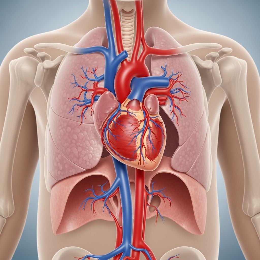
Congenital heart defects like levo-transposition of the great arteries (L-TGA), also known as congenitally corrected transposition, occur when the heart’s major blood vessels are positioned abnormally. In L-TGA, the pulmonary artery and aorta are swapped, but the ventricles are also inverted, allowing somewhat corrected blood flow initially. This condition affects about 0.05% of newborns and demands early detection and intervention to prevent complications.
Understanding the Anatomy of L-TGA
The heart in L-TGA features the morphological right ventricle pumping blood to the body via the aorta, while the left ventricle handles pulmonary circulation. This double switch creates a functional correction but burdens the right ventricle, which is not designed for systemic pressures long-term. Over time, this leads to ventricular strain, valve issues, and conduction abnormalities.
- Key anatomical features: Inverted ventricles, transposed great arteries, frequent atrioventricular block risks.
- Blood flow pathway: Systemic venous blood enters right atrium, crosses to morphological left ventricle, then pulmonary artery; oxygenated blood from lungs to left atrium, morphological right ventricle, aorta.
Associated anomalies like ventricular septal defects (VSD) or pulmonary stenosis occur in over 80% of cases, complicating the clinical picture.
Recognizing Symptoms in Infants and Children
Symptoms vary by associated defects and age. Many infants remain asymptomatic initially due to the ‘corrected’ circulation, but issues emerge as the heart faces stress.
| Age Group | Common Symptoms |
|---|---|
| Newborns/Infants | Cyanosis, heart failure signs like poor feeding, rapid breathing, failure to thrive. |
| Children/Toddlers | Fatigue during play, recurrent infections, growth delays. |
| Older Children | Arrhythmias, exercise intolerance, chest pain. |
In neonates, severe cases present with heart failure within weeks; milder forms may go undetected until school age when arrhythmias or tricuspid regurgitation surface.
Diagnostic Approaches for L-TGA
Diagnosis often starts prenatally via fetal echocardiography, identifying the transposed vessels. Postnatally, tools include:
- Echocardiography: Gold standard, visualizes anatomy, function, and associated defects.
- ECG: Reveals conduction delays or complete heart block in up to 2% at birth, rising to 50% lifetime risk.
- Chest X-ray: Shows cardiomegaly or increased pulmonary markings.
- MRI/Cardiac Catheterization: For detailed anatomy in complex cases.
Early diagnosis is critical; undiagnosed L-TGA risks sudden heart block or systemic ventricular failure.
Treatment Options and Surgical Interventions
Management is tailored to symptoms and anatomy. Asymptomatic patients undergo monitoring; symptomatic ones require intervention.
Medical Management
Medications control heart failure (diuretics, ACE inhibitors), arrhythmias (beta-blockers), and prevent clots. Pacemakers address bradyarrhythmias.
Surgical Strategies
Surgery aims to restore normal anatomy or physiology:
- VSD Closure/Pulmonary Banding: For infants with VSD, to train the left ventricle.
- Double Switch Operation: Atrial and arterial switches to place left ventricle systemic, ideal if done early.
- Valve Replacement: For tricuspid or pulmonary regurgitation.
Timing varies; neonatal repairs for severe cases, later for stable patients. Post-op, lifelong cardiology follow-up is essential.
Long-Term Outlook and Prognosis
With advances, many reach adulthood, but challenges persist. Systemic right ventricle fails in 20-30% by age 40; arrhythmias affect 50%. Regular imaging monitors function; transplants may be needed in end-stage failure.
- Positive factors: Early surgery, no significant residuals.
- Risks: Heart block (1-2% yearly), endocarditis, sudden death.
Multidisciplinary care improves survival to over 80% into adulthood.
Daily Life and Family Guidance
Parents play a vital role. Encourage low-infection environments, prompt illness reporting. Activity: Avoid competitive sports initially; supervised play as tolerated.
- Nutrition: Heart failure diets if needed; growth monitoring.
- Medications: Strict adherence.
- Schooling: IEPs for fatigue-related accommodations.
Psychosocial support via groups like Children’s Heart Foundation aids coping.
Potential Complications to Monitor
Beyond failure, watch for:
- Protein-losing enteropathy from high venous pressures.
- Liver congestion.
- Neurodevelopmental delays from cyanosis or surgery.
Annual Holter monitors, echoes detect issues early.
Frequently Asked Questions (FAQs)
What is the difference between D-TGA and L-TGA?
D-TGA requires urgent arterial switch; L-TGA is ‘corrected’ but risks later failure.
Can children with L-TGA lead normal lives?
Many do with monitoring; restrictions on intense sports apply.
Is L-TGA hereditary?
Rarely; mostly sporadic, screen family if multiple defects.
When is surgery typically performed?
Depends on symptoms; from infancy for severe, later for mild.
What is the survival rate post-double switch?
Over 90% short-term; long-term data evolving.
References
- Pediatric Gastroesophageal Reflux Disease — University of Rochester Medical Center. 2023. https://www.urmc.rochester.edu/encyclopedia/content?ContentTypeID=90&ContentID=P01994
- Gastroesophageal Reflux Disease (GERD) — Children’s Hospital of Philadelphia. 2024. https://www.chop.edu/conditions-diseases/gastroesophageal-reflux-ger
- Gastroesophageal Reflux in Children — Stanford Children’s Health. 2023. https://www.stanfordchildrens.org/en/topic/default?id=gastroesophageal-reflux-in-children-90-P02364
- Reflux in Children — MedlinePlus, U.S. National Library of Medicine. 2025. https://medlineplus.gov/refluxinchildren.html
- Is gastroesophageal reflux disease really a life-long disease — PubMed (NCBI). 2006. https://pubmed.ncbi.nlm.nih.gov/16542297/