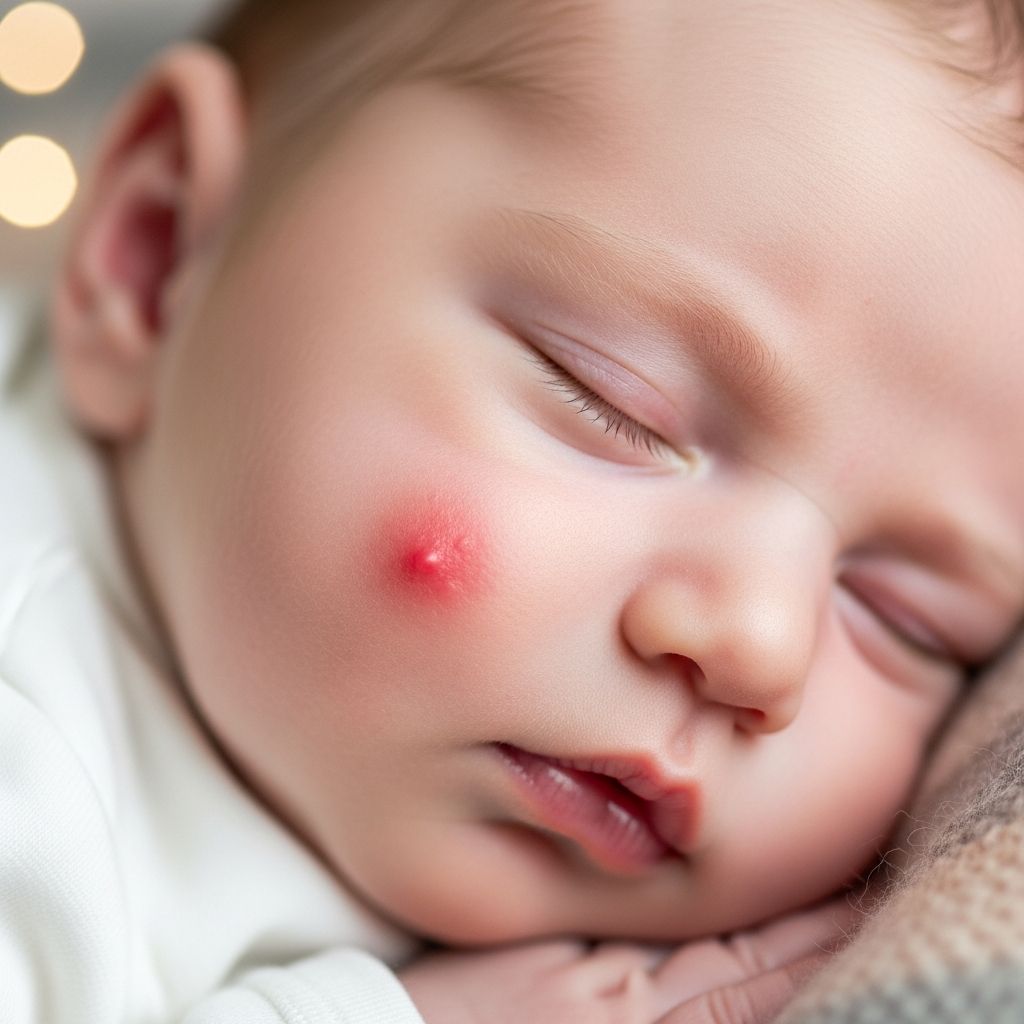
Infantile hemangiomas represent the most prevalent type of benign vascular tumors observed in early childhood, affecting a notable percentage of newborns and infants. These growths, often likened to strawberry marks due to their vivid coloration and textured appearance, emerge from clusters of extra blood vessels and follow a predictable lifecycle of proliferation followed by gradual fading.
Defining Infantile Hemangiomas
These non-cancerous lesions distinguish themselves through a unique developmental trajectory. Unlike permanent vascular birthmarks, infantile hemangiomas typically remain inconspicuous at birth, manifesting as faint patches or subtle discolorations shortly thereafter. Composed of densely packed, immature blood vessels, they can protrude as spongy, rubbery masses on the skin’s surface or lurk deeper beneath, presenting as bluish or flesh-toned swellings.
Prevalence estimates indicate that roughly 4-5% of infants develop at least one such hemangioma, with some experiencing multiples. Their commonality underscores the importance of parental awareness, as most resolve without intervention, yet select cases demand vigilant oversight.
Risk Factors and Underlying Causes
The precise etiology of infantile hemangiomas eludes full comprehension, though research points to disrupted blood vessel formation during fetal development. Hypoxia-related stressors, potentially stemming from placental issues or maternal conditions like preeclampsia, may trigger molecular pathways involving hypoxia-inducible factor-1-alpha (HIF-1α), glucose transporter-1 (GLUT-1), and vascular endothelial growth factor (VEGF-A).
Key demographic and perinatal risk factors include:
- Female infants: Girls face a higher likelihood, comprising the majority of cases.
- Prematurity and low birth weight: Babies born early or small exhibit elevated risk, possibly linked to hypoxic environments.
- Caucasian ethnicity: More frequent in white children compared to other groups.
- Multiples: Twins or triplets show increased incidence.
No direct ties exist to parental diet, genetics, or environmental exposures post-birth, alleviating common parental guilt.
Characteristic Growth Phases
Infantile hemangiomas evolve through distinct stages, aiding diagnosis and management planning:
| Phase | Timeline | Description |
|---|---|---|
| Precursor | Birth to 2 weeks | Faint red patch, telangiectasia, or pale area; minimal visibility. |
| Proliferation | 2 weeks to 9-12 months | Rapid expansion; bright red, raised, spongy texture; may ulcerate. |
| Plateau | 9-12 months | Growth halts; softening begins. |
| Involution | 1-10 years | Shrinkage; color fades to gray, vessels regress; 50% gone by age 5, 90% by 9. |
This cyclical pattern, marked by GLUT-1 positivity, differentiates them from other vascular anomalies.
Types Based on Depth and Extent
Hemangiomas classify by location and distribution:
- Superficial: Raised, bright red ‘strawberry’ marks on skin surface; most common.
- Deep: Subdermal, blue-purple, less defined borders.
- Mixed: Blend of superficial and deep features.
- Localized: Focal, round/oval patches.
- Segmental: Larger (>5cm), plaque-like, following developmental segments; higher complication risk.
Internal sites, notably liver (10-20% of cases), warrant imaging if suspected.
Potential Complications and Warning Signs
While largely harmless, certain hemangiomas pose risks:
- Ulceration: Painful sores from friction; common in proliferating phase.
- Functional impairment: Obstructing eyes, nose, mouth, or genitals.
- High-output heart failure: Rare in massive liver hemangiomas.
- Endocrine issues: Hypothyroidism from liver tumors overproducing type 3 iodothyronine deiodinase.
- Infection: Secondary to ulceration.
Segmental facial hemangiomas signal syndromes like PHACE(S) (posterior fossa anomalies, arterial issues, cardiac defects, eye abnormalities, sternal clefts), LUMBAR (lower body hemangiomas with urogenital anomalies), or PELVIS/SACRAL (genitourinary/spinal defects).
When to Consult a Specialist
Seek prompt evaluation for:
- Periocular growth risking vision.
- Airway or feeding interference.
- Segmental facial lesions (>5cm).
- Lumbosacral involvement suggesting spinal issues.
- Ulceration, bleeding, or rapid growth.
- Multiple or hepatic suspicion (abdominal mass).
Diagnosis relies on clinical exam; biopsy rarely needed due to GLUT-1 marker. Ultrasound/Doppler assesses depth/vessels; MRI for complex cases.
Management and Treatment Strategies
Observation suits most, given spontaneous regression. Active intervention targets complications:
- Topical beta-blockers: Timolol for small, superficial lesions.
- Oral/systemic beta-blockers: Propranolol first-line for problematic cases; shrinks via vasoconstriction/apoptosis.
- Pulsed dye laser: For residual telangiectasias post-involution.
- Surgery: Rare, for residual deformity or fibrosis.
Propranolol dosing starts low (1mg/kg/day), monitored for bradycardia/hypoglycemia. Multidisciplinary teams (dermatology, ophthalmology) optimize outcomes.
Long-Term Prognosis and Scar Management
Over 90% fully regress by adolescence, often leaving atrophic skin, telangiectasias, or fibrofatty residue. Cosmetic lasers address residuals. Early treatment minimizes scarring.
FAQs
Will my baby’s hemangioma disappear completely?
Most fade significantly by school age, though minor skin changes may persist; treatment enhances outcomes.
Is a hemangioma dangerous?
Benign and non-cancerous; risks arise from location/size, not malignancy.
Can hemangiomas be prevented?
No known prevention; risk factors like prematurity are not modifiable.
How soon does treatment work?
Beta-blockers show 30-50% volume reduction in weeks; full effects in months.
Should I worry about multiple hemangiomas?
Common and usually benign; screen for liver involvement if >5-6.
References
- Infantile Hemangiomas: An Updated Review on Risk Factors … — PMC/NCBI. 2018-03-15. https://pmc.ncbi.nlm.nih.gov/articles/PMC5839165/
- Hemangioma – Symptoms and causes — Mayo Clinic. 2023-11-01. https://www.mayoclinic.org/diseases-conditions/hemangioma/symptoms-causes/syc-20352334
- Infantile Hemangioma — Children’s Hospital Colorado. 2024-05-20. https://www.childrenscolorado.org/conditions-and-advice/conditions-and-symptoms/conditions/infantile-hemangioma/
- Infantile Hemangiomas — Children’s Wisconsin. 2023-08-10. https://childrenswi.org/find-care/birthmarks-and-vascular-anomalies/conditions/infantile-hemangiomas
- Infantile Hemangioma | Fact Sheets — Yale Medicine. 2024-02-14. https://www.yalemedicine.org/conditions/infantile-hemangioma
- A Parents’ Guide to Infantile Hemangioma — Hemangeol. 2023-12-05. https://hemangeol.com/a-parents-guide-to-infantile-hemangioma-everything-you-need-to-know/
- Infantile hemangioma — UCSF Benioff Children’s Hospitals. 2024-01-22. https://www.ucsfbenioffchildrens.org/conditions/infantile-hemangioma