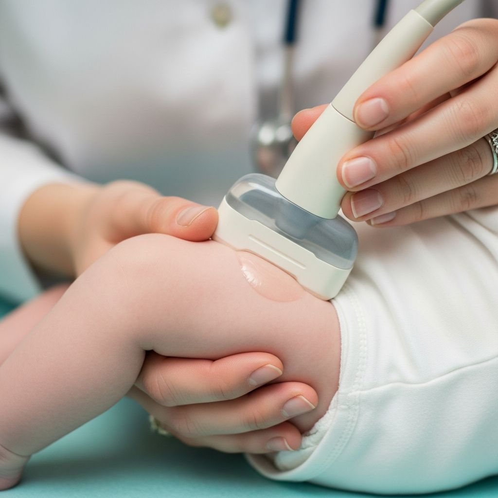
Infant hip ultrasound is a safe, non-invasive imaging test that uses high-frequency sound waves to evaluate the structure and stability of a baby’s hip joints. This procedure is particularly valuable for identifying **developmental dysplasia of the hip (DDH)**, a condition where the hip socket does not fully form or the femoral head does not fit properly, potentially leading to long-term mobility issues if untreated. Early detection through ultrasound allows for timely intervention, often preventing the need for surgery.
Understanding Developmental Dysplasia of the Hip (DDH)
DDH occurs when the ball-shaped top of the thigh bone (femoral head) does not sit securely in the cup-shaped socket (acetabulum) of the pelvis. In mild cases, the head may wobble within the socket; in severe instances, it dislocates completely. This condition affects approximately 1 in 1,000 infants, with girls, first-born children, and those born breech being at higher risk.
During fetal development, the hip joint is held together by soft cartilage rather than bone, making it susceptible to instability. Factors like limited uterine space (e.g., breech position) or family history can contribute. If undetected, DDH may cause uneven leg lengths, limping, or chronic pain later in life.
When Is a Hip Ultrasound Recommended?
Pediatricians routinely screen newborns for hip issues during well-baby visits using physical maneuvers like the Barlow and Ortolani tests, which check for hip instability by gently moving the thighs. An ultrasound is indicated if:
- The baby was in breech position at birth.
- There is a family history of DDH.
- Physical exam reveals limited thigh abduction, leg length differences, or asymmetrical skin folds.
- The infant has neuromuscular conditions or was exposed to oligohydramnios.
Guidelines from the American Institute of Ultrasound in Medicine (AIUM) recommend ultrasound for high-risk infants after 4-6 weeks of age, as earlier scans may show normal immaturity. For low-risk babies, screening is typically not needed unless physical findings suggest concern.
Preparing Your Baby for the Ultrasound
Preparation is minimal, making this test parent-friendly. Key tips include:
- Schedule when your baby is likely to be calm, such as after feeding or naptime.
- Bring comfort items like a bottle, pacifier, blanket, or favorite toy.
- Arrive 15 minutes early with any referral paperwork.
- Plan for only essential family members; additional children may not be allowed in the exam room to minimize distractions.
No fasting or sedation is required, and the baby should be at least 6 weeks old for accurate results. Dress your infant in loose clothing for easy access to the hips.
Step-by-Step Procedure Overview
The exam typically lasts 15-20 minutes and is performed by a trained sonographer or radiologist in a quiet, dimly lit room. Here’s what to expect:
- Positioning: Your baby lies on their back or side on a padded table, often swaddled for comfort. Hips may be flexed in a ‘frog-leg’ position.
- Gel Application: Warm, water-based gel is applied to the hip area—no greasy residue.
- Scanning: A high-frequency linear transducer (probe) is pressed gently against each hip. Images are captured in coronal (front-to-back) and transverse (side-to-side) planes, including neutral, stressed (stress test for stability), and dynamic views.
- Measurements: Key metrics like the alpha angle (acetabular roof angle) and beta angle (cartilage coverage) are assessed. An alpha angle under 60 degrees post-3 months may indicate dysplasia.
- Bilateral Check: Both hips are scanned for comparison.
Parents can soothe the baby by talking, singing, or maintaining eye contact throughout.
Technical Aspects of the Scan
Sonographers use a 9-15 MHz linear probe for optimal soft tissue visualization, focusing on landmarks like the ilium, ischium, femoral head, and labrum. Standard views include:
| View | Purpose | Key Landmarks |
|---|---|---|
| Coronal Standard | Assess acetabular depth | Ilium (horizontal), femoral head centered |
| Coronal with Stress | Check stability | Labrum, femoral metaphysis under pressure |
| Transverse | Evaluate femoral head position | Ischium, femoral shaft |
Dynamic maneuvers simulate pediatrician exams to detect subluxation in real-time.
Interpreting the Results
Results are classified using the Graf method:
- Type I: Normal (alpha >60°).
- Type II: Immature (monitor).
- Type III/IV: Dysplastic/dislocated (treatment needed).
Preliminary findings may be discussed immediately, with a formal radiologist report sent to your doctor within days. Normal hips show the femoral head centered in a deep acetabulum; abnormal ones reveal shallow sockets or displacement.
Risks, Safety, and Comfort Measures
Ultrasound poses no radiation risk, unlike X-rays, making it ideal for infants. The probe pressure is minimal, and gel is hypoallergenic. Babies may fuss due to novelty, but distress is rare. No known adverse effects from diagnostic ultrasound exist per AIUM guidelines.
Treatment Options if DDH is Detected
Early intervention yields excellent outcomes (95% success without surgery). Common approaches:
- Pavlik Harness: A soft brace worn 24/7 for 6-12 weeks to position hips correctly.
- Closed Reduction: Manual repositioning under anesthesia for partial dislocations.
- Surgery: Rare, for persistent cases post-6 months.
Follow-up ultrasounds monitor progress every 4-6 weeks. Most children achieve normal development with prompt care.
FAQs
At what age is hip ultrasound most reliable?
After 4-6 weeks, when physiological immaturity resolves, up to 6 months before bones ossify.
Does the test hurt my baby?
No, it’s painless; only mild probe pressure and gel sensation.
How accurate is infant hip ultrasound?
Highly accurate for DDH detection, with specificity up to 98% in studies.
What if results are borderline?
Repeat scans or observation may be advised, as hips can mature naturally.
Can I hold my baby during the scan?
Often yes, especially if assisting with positioning.
Long-Term Outlook and Prevention Tips
With screening, 90-95% of DDH cases are caught early, averting complications like osteoarthritis. Prevention includes avoiding tight swaddling (keep hips flexible) and using wide-carrier slings. Regular pediatric checkups remain key.
This guide empowers parents to navigate hip ultrasounds confidently, ensuring optimal hip health for their little ones.
References
- Infant Hip Ultrasound – Imaging and Radiology — Henry Ford Health. 2023. https://www.henryford.com/Services/Imaging/Services/Ultrasound/Infant-Hip
- Ultrasound: Infant Hip — Nemours KidsHealth. 2023. https://kidshealth.org/en/parents/ultrasound-hip.html
- Hip Ultrasound — YouTube (Educational Video). 2020. https://www.youtube.com/watch?v=SoUWHKbBHzQ
- Pediatric Hip — American College of Emergency Physicians (ACEP). 2023. https://www.acep.org/sonoguide/advanced/pediatric-hip
- AIUM Practice Parameter for Ultrasound of the Infant Hip — American Institute of Ultrasound in Medicine. 2020. https://www.aium.org/docs/default-source/resources/guidelines/hip.pdf?sfvrsn=9b73e0da_1
- Developmental Dysplasia of the Hip – Ultrasound — Radiology Assistant. 2023. https://radiologyassistant.nl/pediatrics/hip/developmental-dysplasia-of-the-hip-ultrasound