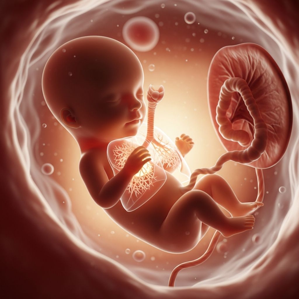
The human respiratory system begins forming in the earliest weeks of pregnancy, evolving through distinct phases to equip the fetus for independent breathing after birth. While fetuses rely on the placenta for oxygen, their lungs undergo complex maturation, including practice breaths of amniotic fluid.
Early Foundations: Embryonic Origins of the Lungs
Lung development initiates around week 4 of gestation when endodermal tissue from the foregut protrudes to form the lung bud, which splits into the laryngotracheal bud. This structure elongates, with the proximal part maturing into the trachea and the distal end branching into bronchial buds—three on the right (forming upper, middle, and lower lobes) and two on the left (upper and lower lobes).
During the embryonic stage (weeks 4-7), these buds establish the basic airway framework. By week 5, the fetus is merely 2 mm long, yet major organ primordia emerge, including the respiratory groove called the sulcus laryngotrachealis in the ventral pharynx.
- Key milestone: Fusion of olfactory pits with pharyngeal endoderm sets the stage for upper airways.
- Branching begins: Repeated divisions create a tree-like pattern, mimicking adult lung architecture.
This phase is critical; disruptions can lead to congenital anomalies like tracheal agenesis.
Airway Branching: Pseudoglandular Phase
From weeks 5-17 (or 7-16 in some models), the pseudoglandular stage focuses on airway formation. Bronchial buds branch up to 20 times, producing segmental bronchi by week 16. Bronchi lumens widen from week 13, and respiratory bronchioles appear.
By the end of this period, all major conducting airways—trachea, bronchi, and bronchioles—are in place, though gas exchange units are absent. The right lung grows larger to accommodate the heart’s position on the left.
| Week Range | Developments | Clinical Note |
|---|---|---|
| 7-13 | Bronchial branching, lumen expansion | Foundation for ventilation pathways |
| 13-16 | Respiratory bronchioles form | All airway structures present |
No gas exchange occurs yet; lungs produce amniotic fluid, aiding fetal urine output and swallowing practice.
Vascularization and Cell Differentiation: Canalicular Stage
Weeks 16-26 mark the canalicular phase, where respiratory bronchioles develop further with extensive blood vessel growth. Alveolar ducts and precursors emerge around week 19, and epithelial cells differentiate into type I (for gas diffusion) and type II pneumocytes (surfactant producers).
Type II cells start secreting pulmonary surfactant by week 20, a phospholipid mix reducing surface tension to prevent alveolar collapse post-birth. Vascularization creates a large gas exchange surface.
- Flattening epithelium: Prepares thin barrier for oxygen-carbon dioxide transfer.
- Capillary proximity: Blood vessels align closely with future alveoli.
This stage’s immaturity in preemies heightens respiratory distress risk due to low surfactant.
Expansion and Maturation: Saccular and Alveolar Phases
The saccular phase (weeks 24-birth) sees saccules (early air sac precursors) enlarge at bronchiolar ends. Surfactant production ramps up, achieving adequate levels by month 8 for lung compliance. Week 28 is pivotal: sufficient maturation allows most 28-week preemies to breathe independently, though full function lags.
From week 36 into childhood, the alveolar phase unfolds. True alveoli form, multiplying from 20-50 million at birth to full capacity by age 8. Lungs expand with body growth, filling the rib cage.
Post-birth, thoracic compression expels lung fluid, and the first cry inflates alveoli with air.
The Mystery of Fetal Breathing Movements
Around weeks 20-21, fetuses exhibit breathing-like motions: diaphragm and intercostal contractions draw in amniotic fluid, exhaling it with surfactant and mucus. These intermittent episodes—frequent then absent—tone respiratory muscles and promote lung expansion.
Though oxygen comes via placenta, these movements are vital for neuromuscular training and surfactant distribution. Ultrasound detects them as rhythmic abdominal undulations, signaling healthy development.
- Frequency: Peaks in low-oxygen states or REM sleep-like phases.
- Benefits: Prevents atelectasis (lung collapse), aids fluid circulation.
Surfactant’s Lifesaving Role
Pulmonary surfactant, produced by type II cells, is crucial from the canalicular stage. It lowers alveolar surface tension, enabling expansion and stability. Insufficient levels cause infant respiratory distress syndrome (IRDS) in preemies.
By term, surfactant ensures smooth transition to air breathing. Antenatal steroids accelerate production in threatened preterm labor.
Risks of Prematurity on Lung Function
Preterm birth interrupts development: before week 24, survival is rare without intervention; post-28 weeks, viability rises, but ventilator support may cause bronchopulmonary dysplasia. Ongoing alveolar growth post-birth underscores neonatal care’s delicacy.
| Gestational Age | Lung Maturity | Survival Odds |
|---|---|---|
| <24 weeks | Canalicular, minimal surfactant | Low, high distress risk |
| 24-27 weeks | Early saccular | Improving with NICU |
| 28+ weeks | Adequate surfactant | Good, independent breathing |
Transition to Air Breathing at Birth
Birth triggers profound changes: cord clamping halts placental gas exchange, prompting chemoreceptors to initiate gasps. Lung fluid clearance via lymphatics and pulmonary circulation shift completes the switch. Surfactant prevents collapse during tidal breathing.
First breath overcomes high resistance, inflating fluid-filled lungs; subsequent breaths ease as compliance improves.
Frequently Asked Questions
Do fetuses breathe in the womb?
Yes, fetuses practice breathing by inhaling and exhaling amniotic fluid starting around week 20, strengthening muscles for birth.
When does surfactant production begin?
Type II cells start producing surfactant around week 20, with levels sufficient by the eighth month.
Can preemies breathe on their own?
From week 28, many can with adequate surfactant; earlier births often require support.
How many alveoli does a newborn have?
Newborns have 20-50 million alveoli, increasing to full numbers by age 8.
What causes fetal breathing movements?
They result from diaphragm contractions, possibly triggered by low oxygen or sleep cycles, aiding lung preparation.
Monitoring Fetal Lung Health
Ultrasounds visualize breathing movements; lecithin-sphingomyelin ratio in amniotic fluid assesses surfactant maturity. Biophysical profiles include breathing as a wellness indicator.
Expecting parents should discuss preterm risks with providers; avoiding smoking supports optimal development.
References
- Embryonic Development of the Respiratory System — Oregon State University Open Textbook. 2023. https://open.oregonstate.education/anatomy2e/chapter/embryonic-development-respiratory-system/
- Development of the respiratory system — Wikipedia (informed by primary sources). 2023. https://en.wikipedia.org/wiki/Development_of_the_respiratory_system
- Fetal Lung Development Stages And Phases — A.T. Respiratory Lectures. 2023. https://atrespiratorylectures.com/blog/posts/fetal-lung-development-stages-and-phases-overview-and-study-guide
- Respiratory System and Disorders — OER Collective (CAUL). 2023. https://oercollective.caul.edu.au/neonatal-care-essentials/chapter/respiratory-system-and-disorders/
- Fetal Development: Stages of Growth — Cleveland Clinic. 2023. https://my.clevelandclinic.org/health/articles/7247-fetal-development-stages-of-growth