Fibular hemimelia is a rare condition that affects the bones of the lower leg and foot, often leading to a shorter leg and differences in the shape or function of the ankle and foot. Although the diagnosis can feel overwhelming, many children with this condition grow up to walk, run, play, and live active lives with the right care and support.
What Is Fibular Hemimelia?
Fibular hemimelia is a congenital limb difference in which part or all of the fibula, the thinner of the two bones in the lower leg, does not form fully before birth. The tibia (the larger shinbone), knee, ankle, and foot may also develop differently, leading to a leg that is shorter and sometimes curved, with possible differences in the number or shape of the toes.
The condition typically affects one leg (unilateral), but in a smaller number of children, both legs are involved (bilateral). Fibular hemimelia is one of the more common causes of significant leg length discrepancy in childhood, even though it is still considered a rare condition overall.
How the Lower Leg Is Normally Built
To understand fibular hemimelia, it helps to know the basic structure of the lower leg. From the knee to the ankle, there are two main bones: the tibia on the inner side and the fibula on the outer side.
- Tibia: Bears most of the body’s weight and forms the main part of the knee and ankle joints.
- Fibula: Supports the outer side of the leg, helps stabilize the ankle, and provides attachment points for important muscles and ligaments.
- Foot bones: Many small bones form the arch and toes, allowing balance and movement.
When the fibula is very short or absent, the way forces travel through the leg changes. This can affect alignment of the knee, position of the ankle, and the way the foot contacts the ground.
Common Features and Signs
Fibular hemimelia can look different from one child to another, ranging from mild differences to more obvious changes in leg length and foot shape. Some features are visible at birth, while others become more noticeable as the child grows and begins to stand or walk.
- One leg shorter than the other at birth or early in infancy.
- A thinner or slightly bowed lower leg on the affected side.
- Differences in the ankle, such as the foot appearing higher in the front or tilted outward.
- Fewer toes on the affected foot, or toes that are joined or shaped differently.
- Changes higher up the leg, such as a shallow hip socket or differences in knee stability.
As the child grows, the shorter leg usually continues to grow more slowly, which can increase the difference in length. Parents may notice limping, frequent tripping, or the child preferring to stand on the longer leg.
Why Does Fibular Hemimelia Happen?
Fibular hemimelia develops very early in pregnancy, when the baby’s limb buds are forming and the major bones are beginning to take shape. For reasons that are not fully understood, the cells that should form the fibula and nearby structures do not develop in the usual way.
In most families, fibular hemimelia is not inherited and is not caused by anything the parents did or did not do. Researchers suspect that a combination of genetic influences and local growth disturbances during limb development may play a role, but no single cause has been identified for most children.
How Doctors Diagnose the Condition
Diagnosis usually begins with a physical exam soon after birth, when the shorter leg, unusual foot position, or missing toes are first noticed. The healthcare team will assess how the hip, knee, and ankle move and how stable these joints are.
Imaging tests help build a clearer picture of the bones and joints:
- X-rays: Show the length and shape of the tibia and fibula, the alignment of the knee and ankle, and the number and position of foot bones.
- Ultrasound or MRI: May be used to evaluate cartilage, ligaments, and the hip joint, especially when planning surgery.
- Growth predictions: Specialized charts and computer models help estimate how large the leg length difference may become in later childhood or adolescence.
Sometimes, fibular hemimelia occurs along with other limb differences, such as congenital femoral deficiency (differences in the thighbone), so the entire lower limb is carefully evaluated.
Severity Levels and Classifications
Because fibular hemimelia varies widely, orthopedic specialists often use classification systems to describe how much of the fibula is missing and how the ankle and foot are affected. These systems help guide treatment planning but are primarily used by the medical team rather than families in day-to-day life.
In general, the condition can be thought of across a spectrum:
- Milder forms: Smaller leg length difference, a mostly stable ankle, and a foot with near-normal structure.
- Moderate forms: Noticeable leg length difference, partial fibula absence, and a foot with fewer or misshapen toes.
- More severe forms: Near or complete absence of the fibula, major ankle instability or stiffness, and a significantly smaller or malformed foot.
Overview of Fibular Hemimelia Features
| Aspect | Milder Presentation | More Severe Presentation |
|---|---|---|
| Leg length | Small difference, often a few centimeters | Large difference, may be several inches |
| Fibula | Partially shortened but present | Almost or completely absent |
| Ankle joint | Reasonably stable with some deformity | Significantly unstable or stiff |
| Foot and toes | Near-normal size; minor toe differences | Smaller foot; fewer or fused toes |
Treatment Goals and Big-Picture Planning
The central goal of treatment is to help the child stand, walk, and move as comfortably and safely as possible, with legs that are as equal in length as is practical. Decisions are tailored to the individual child and are revisited over time as the child grows and goals change.
Families typically work with a multidisciplinary team that may include pediatric orthopedic surgeons, physical therapists, prosthetists (for artificial limbs), nurses, social workers, and psychologists. This team approach supports both the child’s physical needs and the family’s emotional and practical concerns.
Non-Surgical Options
In infancy and early childhood, many children benefit from nonsurgical supports to improve alignment and function while long-term plans are being made.
- Shoe lifts or inserts: Used to help even out leg length differences that are mild or during the years between surgeries.
- Braces or orthoses: Devices that support the ankle and foot, create a more stable base for standing, and improve walking patterns.
- Physical therapy: Exercises to build strength, flexibility, and balance, and to encourage symmetrical use of both legs.
Even when surgery is planned, these conservative measures can make daily life safer and more comfortable and may reduce strain on the spine and hips.
Surgical Limb Lengthening and Reconstruction
One major treatment path is limb reconstruction, in which surgeons try to preserve the child’s foot and gradually lengthen the shorter bones. This approach often involves multiple stages spread out over several years.
Key Elements of Reconstruction
- Correcting deformities: Procedures to straighten the tibia, realign the ankle, or reshape the foot to create a more plantigrade (flat, weight-bearing) position.
- Limb lengthening: Specialized devices slowly separate the bone after a controlled cut, allowing new bone to form in the gap while soft tissues stretch.
- Joint stabilization: Surgery to tighten or reconstruct ligaments or, in some cases, fuse certain joints for improved stability.
Limb lengthening is a complex process that requires close follow-up, daily adjustments of the device, and consistent physical therapy. Families should be prepared for an intensive but potentially very rewarding experience that can significantly reduce the final leg length difference.
Amputation and Prosthetic Limb Care
For some children, especially those with very small or severely deformed feet and ankles, amputation of the foot or part of the leg followed by fitting with a prosthetic limb offers the best long-term function. This option may allow earlier walking and more predictable leg length as the child grows.
What This Approach Involves
- Planned surgery in early childhood: Often performed within the first year or two of life so the child can learn to walk using a prosthesis from the start.
- Custom prosthetic limb: Designed to match the child’s size and activity level, and adjusted regularly as the child grows.
- Ongoing support: Physical therapy and prosthetic training to help the child move confidently and participate in play and sports.
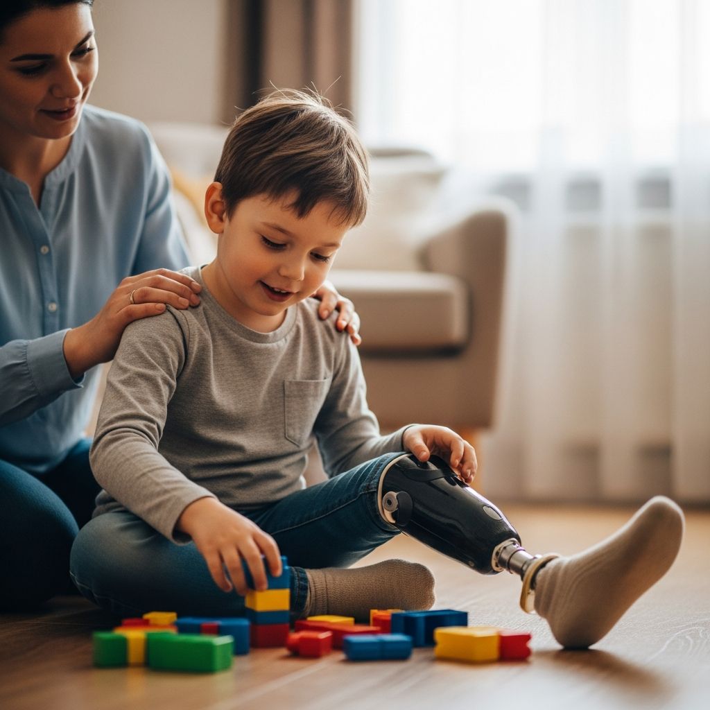
Many children using a prosthesis enjoy high levels of activity, including running, biking, and participating in organized sports. Families considering this path often find it helpful to meet other parents and children who have followed a similar route.
Living Day to Day With Fibular Hemimelia
Beyond medical treatment, families manage many practical issues, from choosing supportive footwear to coordinating school and therapy schedules. Good communication with teachers, coaches, and other caregivers helps ensure that the child’s needs are understood and accommodations are in place when necessary.
- Encourage the child to stay active within comfort and safety limits.
- Teach peers simple, respectful language about limb differences to reduce teasing and confusion.
- Celebrate the child’s abilities and problem-solving skills, not just their medical milestones.
Emotional support is just as important as physical care. Some families benefit from counseling or support groups where children and parents can share their experiences and learn coping strategies.
Long-Term Outlook
With modern orthopedic techniques, most children with fibular hemimelia can expect to walk independently and participate in everyday activities. The specific path—limb lengthening, prosthetic care, or a combination—depends on the details of each child’s anatomy and family preferences.
Regular follow-up is essential, often continuing into adolescence, to monitor growth, joint health, and function. Adjustments to bracing, prostheses, or surgical plans may be needed as the child’s goals evolve, from learning to walk to joining sports or preparing for adult life.
Frequently Asked Questions (FAQs)
Is fibular hemimelia painful for children?
Many infants and young children with fibular hemimelia are not in constant pain, but they may experience discomfort from leg length differences, joint strain, or skin irritation around braces or devices. After surgery or during limb lengthening, temporary pain or soreness is common and is managed with medications, rest, and careful monitoring by the care team.
Can my child play sports?
Many children with fibular hemimelia safely take part in physical education and recreational sports, sometimes with modifications. The specific activities that are appropriate will depend on the child’s strength, balance, and any devices or prostheses they use, so it is important to discuss plans with the orthopedic team and therapist.
Will more than one surgery be needed?
It is common for children with fibular hemimelia to need more than one procedure during childhood and adolescence, especially if limb lengthening is chosen. Each surgery usually has a specific goal, such as improving alignment, stabilizing a joint, or adding length as the child grows.
Does this condition affect life expectancy?
Fibular hemimelia primarily involves the bones and joints of the leg and does not typically change overall life expectancy. The main focus is on maximizing mobility, comfort, and participation in everyday life.
How can our family find support?
Families often find it helpful to connect with national or local limb difference organizations, online communities, or hospital-based parent groups. These resources can provide practical tips, emotional support, and a sense of community with others who understand the challenges and successes of living with fibular hemimelia.
Key Takeaways for Parents
- Fibular hemimelia is a difference in how the lower leg and foot form before birth, leading to a shorter leg and possible changes in the ankle and toes.
- Treatment plans are individualized and may include bracing, shoe lifts, limb lengthening and reconstruction, or amputation with prosthetic fitting.
- With appropriate care, many children with fibular hemimelia lead active, independent lives and participate fully in school, play, and family activities.