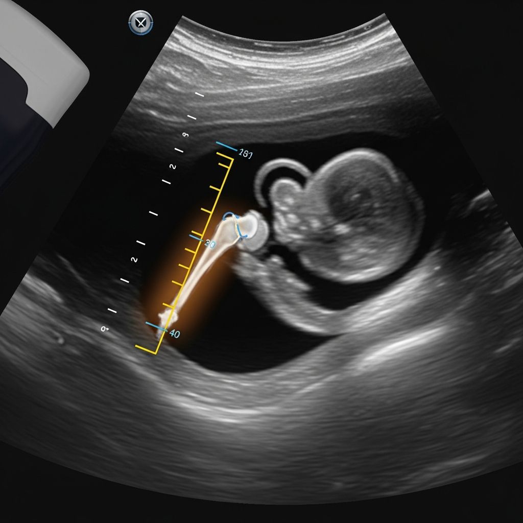
The femur length, often abbreviated as FL on ultrasound reports, represents the measurement of the thigh bone in a developing fetus. This biometric parameter serves as a cornerstone in prenatal care, offering insights into the baby’s growth trajectory, gestational age estimation, and overall skeletal health.
Why Femur Length Matters in Fetal Development
During routine pregnancy scans, sonographers capture several key measurements to evaluate fetal well-being. Among these, femur length stands out because the femur is the longest bone in the human body, making it a reliable indicator of proportional growth. From the second trimester onward, FL helps clinicians confirm if the fetus is developing at an expected pace.
Tracking FL over multiple ultrasounds allows healthcare providers to plot growth curves. Consistent increases in length signal healthy bone formation and nutrient absorption, while deviations can prompt further investigation. This measurement becomes particularly valuable after 14 weeks when the femur is clearly visible on imaging.
How Sonographers Measure Femur Length Accurately
Obtaining a precise FL reading requires skilled technique. The ultrasound probe is positioned to visualize the femur in a longitudinal view, ensuring the entire bone—from the greater trochanter at the hip to the distal condyle at the knee—is straight and unobstructed. Curved or oblique views can lead to underestimation, so standardization is crucial.
Modern ultrasound machines provide real-time calipers for digital measurement, minimizing errors. Factors like fetal position, maternal body habitus, and equipment quality influence accuracy, but trained professionals follow protocols from organizations like the American College of Obstetricians and Gynecologists to ensure reliability.
Normal Femur Length Ranges by Gestational Week
Fetal femur length correlates closely with gestational age, increasing steadily as pregnancy progresses. Charts derived from large population studies define average, percentile ranges, allowing comparisons to norms.
| Gestational Age (Weeks) | Mean FL (cm) | 5th-95th Percentile (cm) |
|---|---|---|
| 20 | 3.35 | 3.0 – 3.7 |
| 24 | 4.40 | 3.9 – 4.9 |
| 28 | 5.30 | 4.7 – 5.9 |
| 32 | 6.10 | 5.5 – 6.7 |
| 36 | 6.85 | 6.2 – 7.5 |
| 40 | 7.50 | 6.8 – 8.2 |
These values are approximate and based on aggregated data; individual variations occur due to genetics and ethnicity. By 37 weeks, a FL around 7.0 cm is typical.
Role of FL in Confirming Gestational Age
Early in the second trimester (12-23 weeks), femur length offers high precision for dating pregnancies, often more accurate than later measurements affected by growth variability. Combined with biparietal diameter (BPD) and abdominal circumference (AC), FL refines due date predictions within days.
If FL aligns with other biometrics, it reassures providers of a well-dated pregnancy. Discrepancies exceeding two weeks may indicate post-term gestation or early growth issues, guiding decisions on induction or monitoring.
Monitoring Fetal Growth Through Serial FL Scans
Isolated FL readings provide snapshots, but serial ultrasounds reveal trends. Growth velocity—the rate of FL increase—should follow established curves. A lag below the 10th percentile across scans suggests intrauterine growth restriction (IUGR), where placental insufficiency limits nutrient delivery.
- Normal growth: Steady percentile tracking between scans.
- Borderline: Crossing percentiles downward, warrants Doppler studies.
- Restricted: Persistent <5th percentile, risking low birth weight.
Conversely, accelerated growth might indicate macrosomia, especially in diabetic pregnancies. Regular FL assessments empower proactive interventions like nutritional counseling or bed rest.
Short Femur Length: Potential Causes and Implications
A femur shorter than the 5th percentile prompts detailed evaluation. In a study of 1373 cases, nearly 50% were isolated findings leading to healthy outcomes, but 28.7% involved additional anomalies.
Common associations include:
- Intrauterine Growth Restriction (IUGR): Often due to placental problems, affecting overall size.
- Constitutional Smallness: Genetically petite babies without pathology.
- Skeletal Dysplasias: Rare bone disorders like achondroplasia, detectable via bowing or rhizomelia.
Isolated short FL (<1st centile, disproportionate to head/AC) heightens genetic testing consideration, such as amniocentesis.
Linking Short FL to Chromosomal and Genetic Risks
Short femur serves as a soft marker for trisomy 21 (Down syndrome), particularly if <5th percentile in the mid-trimester. However, it’s not diagnostic alone—combined with nuchal translucency, nasal bone absence, and serum markers via quadruple test for risk stratification.
Chromosomal issues appeared in 5.5% of short FL cases in large cohorts, underscoring the need for extended anomaly scans and counseling. Other aneuploidies like trisomy 18 may present similarly.
Ratios Involving Femur Length for Deeper Insights
Beyond absolute length, ratios enhance assessment:
| Ratio | Normal Range | Clinical Meaning |
|---|---|---|
| FL/AC | 0.16-0.24 | Low: Asymmetric IUGR; High: Limb overgrowth |
| FL/HC | 0.83-0.92 | Deviation signals disproportionality |
FL/AC ratio flags nutritional imbalances; if low, it suggests abdominal sparing in IUGR. These indices complement estimated fetal weight (EFW) formulas.
Long Femur Length: What It Might Indicate
While short FL garners attention, excessive length (>90th percentile) can signal overgrowth. Common in gestational diabetes, it correlates with macrosomia, elevating cesarean and shoulder dystocia risks. Ethnic norms vary—longer FL in African-American fetuses.
Advanced Imaging and Follow-Up for Abnormal FL
Suspected dysplasias require 3D ultrasound or fetal MRI for bone detail. Doppler velocimetry assesses umbilical artery flow in growth concerns. Multidisciplinary teams, including geneticists, guide management.
Practical Advice for Expectant Parents
Receiving an FL report can spark anxiety. Discuss percentiles with your provider—context matters. Maintain prenatal visits, balanced diet, and avoid smoking to optimize growth. Most short FL cases resolve positively with monitoring.
Frequently Asked Questions About Femur Length
What is a normal femur length at 20 weeks?
Around 3.2-3.5 cm, within 5th-95th percentiles.
Does short femur always mean Down syndrome?
No, it’s a marker, not diagnostic; only 5-10% of cases link to trisomy 21 when isolated.
How often is FL measured in pregnancy?
Typically at 18-22 week anatomy scan and third-trimester growth scans.
Can short FL be due to ethnicity?
Yes, reference charts account for variations; consult tailored norms.
What if FL is below 5th percentile?
Expect serial scans, possible genetic testing, and IUGR surveillance.
This guide equips parents with knowledge for informed discussions. Always rely on your healthcare team’s expertise.
References
- What Does FL Mean on an Ultrasound? Best Guide — Liv Hospital. 2023. https://int.livhospital.com/what-does-fl-mean-on-an-ultrasound/
- Femur Length — Global Ultrasound Institute. 2024. https://globalultrasoundinstitute.com/pocuspedia/femur-length/
- The femur too short? 1373 fetuses with short femur during second trimester screening — PMC (NCBI). 2022-09-06. https://pmc.ncbi.nlm.nih.gov/articles/PMC9470684/
- Presentation: Fetus with an isolated short femur — Genomics Education Programme (NHS). 2023. https://www.genomicseducation.hee.nhs.uk/genotes/in-the-clinic/presentation-fetus-with-an-isolated-short-femur/
- Fetal Biometry: Measurements, Normal Values & Accuracy — Cleveland Clinic. 2024-01-15. https://my.clevelandclinic.org/health/diagnostics/25087-fetal-biometry