Recognizing Ear Canal Narrowing: An Overview for Parents and Caregivers
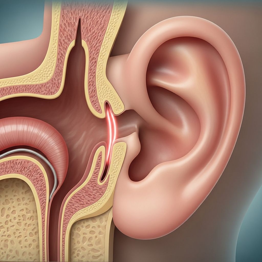
Ear canal stenosis represents a medical condition in which the external auditory canal becomes progressively narrowed, restricting the normal passage of sound vibrations toward the eardrum. This narrowing can occur in isolation or affect both ears simultaneously, and its severity ranges from barely noticeable to profoundly disruptive. The condition may be present from birth or develop later in childhood due to various triggering factors. Understanding this condition is crucial for parents, as early recognition and appropriate intervention can prevent complications and preserve hearing function.
The external ear canal serves as a crucial conduit for sound transmission. When this pathway becomes restricted, even mildly, it can interfere with the normal hearing process and create an environment conducive to infection and debris accumulation. Parents who notice signs of hearing difficulty or repeated ear problems in their children should consult with an ear specialist to determine whether stenosis is present.
Observable Indicators and Clinical Presentations
The manifestations of ear canal stenosis vary considerably depending on the degree of narrowing and whether one or both ears are affected. Mild stenosis may produce no noticeable symptoms whatsoever, discovered only during routine medical examinations. However, as the narrowing becomes more pronounced, several distinctive signs may emerge.
Hearing difficulties represent the most commonly reported symptom when stenosis is moderate to severe. Sound waves cannot penetrate the narrowed canal efficiently, resulting in conductive hearing loss. Children may appear inattentive in classroom settings, request that people speak louder, or show delayed speech development. The degree of hearing impairment correlates directly with the severity of the stenosis.
Recurrent ear infections frequently accompany ear canal stenosis. The narrowed canal creates stagnant pockets where moisture and debris accumulate, providing an ideal breeding ground for bacteria and fungi. External ear infections, medically termed otitis externa, become increasingly common and may prove resistant to standard antibiotic treatments.
Sensation of ear fullness is another frequently reported complaint. Children may describe feeling as though something is blocking their ear or that their ear feels plugged, similar to the sensation experienced during altitude changes. This sensation typically persists and does not resolve spontaneously.
Tinnitus or ear ringing occasionally accompanies stenosis, though this symptom is less common in pediatric patients than in adults. When present, the ringing may be constant or intermittent and may worsen with certain head positions or activities.
Discomfort during specific activities such as chewing or yawning may occur, particularly when stenosis is accompanied by inflammation or infection. Some children report ear pain that intensifies during upper respiratory infections.
Understanding the Origins and Contributing Factors
Ear canal stenosis develops through multiple distinct pathways, each with different risk factors and presentations. Identifying the underlying cause helps guide treatment decisions and may inform preventive strategies.
Congenital Presentations
Some children are born with ear canal stenosis due to abnormal development of the external ear structures during fetal growth. These congenital cases result from genetic factors or developmental anomalies that occur during the critical periods of ear formation. Congenital stenosis may be partial, where a narrowed but patent canal exists, or complete (atresia), where the canal fails to develop entirely. Early detection through newborn hearing screening allows for timely intervention before the child experiences significant developmental delays related to hearing loss.
Acquired Stenosis from Infection and Inflammation
Perhaps the most common pathway to stenosis in children involves repeated ear infections causing scarring and tissue damage. When otitis externa occurs frequently, the inflammatory response can trigger abnormal healing, resulting in progressive narrowing. Chronic fungal infections similarly contribute to tissue changes that lead to canal constriction. The inflammatory cascade initiated by these infections alters normal tissue architecture, depositing scar tissue that gradually reduces canal diameter.
Trauma and Physical Injury
Injuries to the ear canal from aggressive cleaning methods, cotton swabs inserted too deeply, foreign object insertion, or ear piercings can trigger stenosis development. The body’s healing response to these injuries may result in excessive scarring that narrows the canal. Parents should be educated about safe ear cleaning practices to minimize this risk.
Environmental and Moisture-Related Factors
Prolonged exposure to moisture, particularly in children who frequently swim or participate in water sports, can contribute to canal narrowing. Additionally, certain skin conditions such as eczema or psoriasis affecting the ear canal increase inflammation and may promote stenosis development. Some children develop exostoses (bony growths) in response to cold water exposure, which can contribute to canal narrowing, though this mechanism differs from typical stenosis.
Post-Surgical Complications
Children who undergo ear surgery may develop stenosis as a healing complication. Surgical procedures designed to treat other ear conditions occasionally result in unexpected scar tissue formation that restricts the canal. This complication requires vigilant monitoring and may necessitate additional interventions.
Diagnostic Approaches and Medical Evaluation
Accurate diagnosis requires a systematic evaluation combining clinical assessment, hearing evaluation, and imaging studies. An otolaryngologist (ENT specialist) typically coordinates this diagnostic process.
Otoscopic examination allows direct visualization of the ear canal. The specialist can observe the degree of narrowing, assess the canal lining for inflammation or debris, and examine the eardrum for signs of damage or abnormal pressurization.
Audiometric testing measures hearing sensitivity and determines whether conductive hearing loss is present. Age-appropriate testing methods are used based on the child’s developmental level, ranging from behavioral observation in infants to standard audiometry in older children.
Computed tomography (CT) scanning provides detailed three-dimensional images of the ear canal structure, allowing the specialist to assess the extent of narrowing, determine whether bony or soft tissue components predominate, and evaluate middle ear structures for complications such as cholesteatoma. CT imaging becomes particularly important when surgical intervention is being considered.
Tympanometry evaluates middle ear function and can help identify whether fluid or other abnormalities exist behind the eardrum, which sometimes accompany stenosis.
Management Strategies Based on Severity
Treatment approaches vary significantly depending on the severity of stenosis and whether hearing loss or infections are present. A tiered approach allows appropriate matching of intervention intensity to clinical needs.
Conservative Management for Mild Cases
When stenosis is mild and produces no symptoms or minimal hearing impairment, watchful waiting with regular follow-up represents the appropriate initial approach. The child’s hearing is monitored periodically, and the canal is inspected for debris accumulation or infection development.
Preventive measures become important in these cases:
- Avoiding cotton swabs or other objects inserted into the ear canal
- Wearing custom-fitted earplugs during water activities to prevent moisture accumulation
- Maintaining ear hygiene through gentle external cleaning only
- Addressing skin conditions affecting the ear canal promptly
- Treating ear infections aggressively to prevent scarring complications
Pharmacological Interventions
When mild to moderate stenosis is accompanied by inflammation or early infection, topical medications may help manage symptoms and slow progression. Antibiotic ear drops address bacterial colonization, while steroid-containing drops reduce inflammation and may limit scar tissue formation. However, medications cannot reverse established narrowing and serve primarily as adjunctive therapies.
Hearing Aids and Assistive Devices
Children with stenosis-related hearing loss who are not surgical candidates or whose stenosis is not yet severe enough for surgery benefit from hearing aid amplification. Modern pediatric hearing aids provide natural sound amplification while remaining comfortable and cosmetically acceptable for active children. Additionally, bone-anchored hearing aids (BAHA devices) offer an alternative approach that bypasses the external ear canal entirely, transmitting vibrations directly to the inner ear through bone conduction.
Procedural Dilation Techniques
For stenosis causing functional impairment, ear canal dilation represents a non-surgical intervention option. This procedure involves carefully stretching the narrowed canal using specialized instruments under medical supervision. While dilation can provide temporary improvement, restenosis (recurrence of narrowing) frequently occurs within months, particularly when the underlying inflammatory condition persists.
Surgical Reconstruction
Children with severe stenosis causing significant hearing loss or recurrent infections typically require surgical intervention. The primary surgical objective involves creating a wider, patent ear canal (canaloplasty or atresiaplasty for congenital cases). The surgeon may remove excess bone, reshape scar tissue, or utilize skin grafts to increase canal diameter and prevent restenosis.
Surgical success depends on multiple factors, including the surgeon’s experience, the degree of scarring, and the child’s healing characteristics. Post-operative monitoring is essential, as restenosis represents the most common complication, occurring in a significant percentage of surgically treated cases. Some children may require revision procedures to maintain adequate canal patency.
Serious Complications Requiring Attention
Beyond the immediate concerns of hearing loss and infections, stenosis creates conditions favorable for more serious complications that require proactive monitoring.
Cholesteatoma formation represents a particularly important concern. This non-cancerous skin cyst develops in the middle ear or canal when skin and debris become trapped behind a narrowed canal opening. As the cholesteatoma enlarges, it can erode the delicate bones of the middle ear, leading to progressive hearing loss, balance problems, and facial nerve dysfunction. Cholesteatoma requires surgical removal, and the procedure may necessitate reconstruction of damaged ear structures.
Persistent ear infections that resist standard antibiotic therapy can develop when bacteria or fungi become established in the stagnant environment created by stenosis. These chronic infections produce chronic drainage (otorrhea), discomfort, and potential complications including osteomyelitis of the temporal bone.
Long-Term Monitoring and Follow-Up Care
Regardless of the treatment approach selected, children with ear canal stenosis require ongoing specialist oversight. Regular otolaryngological examinations allow assessment of canal patency, early detection of complications, and timely intervention if stenosis progresses or complications develop.
Audiological re-evaluation at appropriate intervals documents any changes in hearing status and guides decisions regarding hearing aid use or surgical intervention. Annual assessments are typical for stable cases, while more frequent monitoring may be necessary following surgical procedures or if symptoms suggest disease progression.
Frequently Asked Questions
Can ear canal stenosis resolve on its own?
Mild stenosis may remain stable without progression, but established narrowing does not spontaneously reverse. Scar tissue that has formed typically persists unless surgically addressed.
At what age should a child with congenital stenosis have surgery?
Surgery for congenital stenosis is typically considered around age five or six, though timing depends on the severity of hearing loss and whether external ear reconstruction is also needed. Earlier intervention may be appropriate in some cases.
What is the success rate for surgical treatment?
While surgery can effectively widen the canal, restenosis (recurrence of narrowing) remains common, occurring in a significant percentage of cases. Some children require revision procedures to maintain adequate patency.
Can ear canal stenosis prevent normal development?
Yes, severe bilateral stenosis causing significant hearing loss during critical language development periods can impact speech and language acquisition. Early identification and intervention are crucial to minimize this risk.
Are there preventive measures to reduce stenosis risk?
While congenital stenosis cannot be prevented, acquired stenosis risk can be reduced through gentle ear cleaning practices, prompt treatment of ear infections, use of protective earplugs during water activities, and management of skin conditions affecting the ear canal.
References
- Ear Canal Stenosis: Signs, Causes, and Treatment Options — Medicover Hospitals. 2024. https://www.medicoverhospitals.in/diseases/ear-canal-stenosis/
- Acquired Stenosis of the External Ear Canal — PubMed Central/National Center for Biotechnology Information. 2023. https://pubmed.ncbi.nlm.nih.gov/37553271/
- Congenital Ear Conditions We Treat: External Auditory Canal Stenosis — UPMC Children’s Hospital. 2024. https://www.chp.edu/our-services/ent/services/congenital-ear/conditions
- Ear Canal Stenosis and Atresia: Surgical Options — Coastal Ear Nose & Throat. 2024. https://coastalearnoseandthroat.com/services/ear-surgery/ear-canal-stenosis-and-atresia/
- Ear Canal Stenosis — Nemours KidsHealth. 2024. https://kidshealth.org/en/parents/ear-canal-stenosis.html
- Ear Canal Exostoses & Stenosis — Stanford Children’s Health. 2024. https://www.stanfordchildrens.org/en/services/hearing-center/conditions/ear-canal.html