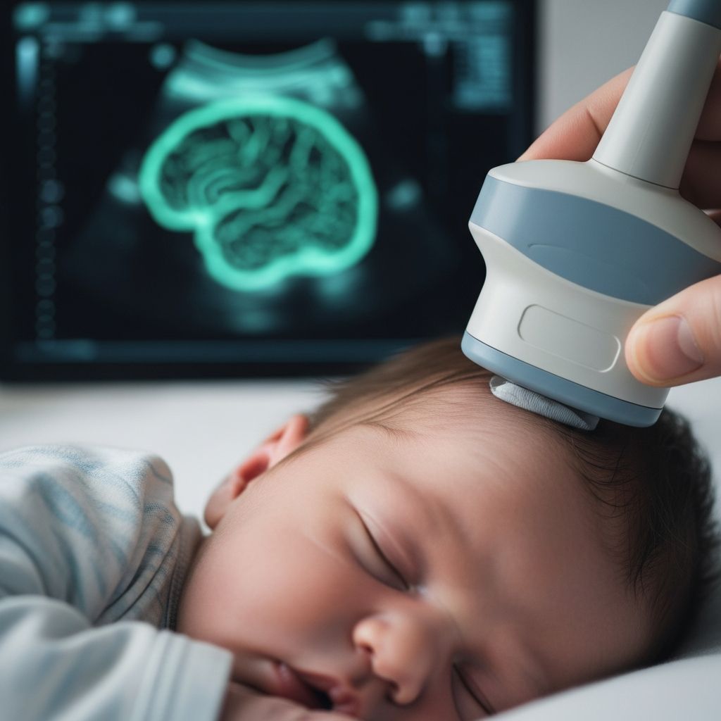
Cranial ultrasound, often called a head ultrasound, is a vital diagnostic tool used primarily on newborns and young infants to examine brain structures safely and effectively. This imaging method employs high-frequency sound waves to create detailed pictures of the brain, accessible through the open fontanelles before skull bones fully fuse.
Understanding the Role of Cranial Ultrasound in Newborn Care
In neonatal intensive care units (NICUs) and pediatric settings, cranial ultrasound serves as the first-line imaging for assessing brain health. It detects issues like bleeding, fluid buildup, or structural anomalies without radiation exposure, making it ideal for fragile preterm babies. Medical professionals rely on it for quick bedside evaluations, allowing repeated scans as needed to monitor progress.
The technique’s portability enables immediate use during critical moments, such as after birth complications or signs of neurological distress. Unlike CT or MRI, which require transport and sedation, ultrasound fits seamlessly into routine newborn checks.
Key Reasons for Performing Head Ultrasounds on Infants
Healthcare providers recommend cranial ultrasounds for various concerns in early infancy. Common indications include:
- Prematurity, to screen for intraventricular hemorrhage (IVH) or periventricular leukomalacia (PVL), which affect white matter around brain ventricles.
- Rapid head growth or macrocephaly, signaling potential hydrocephalus.
- Bulging or unusually large fontanels, which may indicate increased intracranial pressure.
- Neurological symptoms like seizures, poor feeding, or lethargy.
- Suspected infections, trauma, or congenital malformations.
- Follow-up for prenatal findings or post-surgical monitoring.
These scans help diagnose bleeding in brain tissue or ventricles, fluid accumulation (hydrocephalus), tumors, cysts, or meningitis complications. Early detection facilitates timely interventions, improving outcomes.
Technical Aspects: Probes, Frequencies, and Imaging Windows
Performing a high-quality cranial ultrasound requires specialized equipment tailored to infant anatomy. Technicians use small curvilinear or sector probes (7.5-10 MHz for initial scans) to penetrate deeper structures through the anterior fontanelle, the primary acoustic window. Higher-frequency linear probes (9-18 MHz) provide superior resolution for superficial areas like the cortex and extra-axial spaces.
Additional windows include the posterior, temporal, and mastoid fontanelles for comprehensive views, especially of the cerebellum and brainstem. Operators capture coronal and sagittal planes, plus Doppler assessments of vessels like the anterior cerebral artery to evaluate blood flow and resistance index (RI), crucial for conditions like hypoxic-ischemic encephalopathy (HIE).
| Probe Type | Frequency Range | Primary Use |
|---|---|---|
| Curvilinear/Sector | 7.5-10 MHz | Deep brain structures, full scans |
| Linear | 9-18 MHz | Superficial cortex, sinuses, vessels |
Optimal imaging demands symmetric, centered views. Gel is applied after locating the fontanelle, with the infant positioned comfortably—head parallel or perpendicular to the bed.
Step-by-Step Procedure for a Safe Scan
The process is straightforward and gentle, typically lasting 30-60 minutes. No sedation or special preparations are needed, though a pacifier or feeding can soothe the baby. Lights dim to enhance monitor visibility as the sonographer glides the probe over the fontanelle with warm gel.
- Positioning: Baby lies supine; head stabilized gently.
- Gel Application: Water-based gel on probe and skin.
- Scanning: Coronal views through anterior fontanelle, then sagittal, with supplementary windows.
- Doppler: Checks vessel patency and flow velocities.
- Dynamic Tests: Probe compression assesses hydrocephalus type (obstructive vs. communicating).
- Completion: Images reviewed; baby cleaned and comforted.
Parents can stay present, observing the real-time images.
Interpreting Normal and Abnormal Findings
Normal scans show symmetric ventricles, even echotexture, maturing sulci/gyri (smoother in preemies), and patent vessels. Brain maturity evolves rapidly: preterm brains appear primitive at birth but develop gyration by 2 months.
Abnormalities include:
- Germinal Matrix Hemorrhage: Common in preemies; graded by extent, with evolution tracked over time.
- Ventricular Dilatation: Measured via Levene index (falx to lateral ventricle wall); indicates hydrocephalus.
- Parenchymal Damage: Cystic changes or white matter loss from PVL or HIE.
- Masses/Fluid: Cysts, tumors, or extra-axial collections.
Doppler RI helps stage HIE, performed 48-72 hours post-birth.
Safety Profile and Limitations of the Technique
Cranial ultrasound is noninvasive, radiation-free, and sedation-free, posing no known risks even with frequent use. It’s cost-effective and bedside-friendly.
Limitations: Effective only while fontanelles remain open (up to 6-12 months, best <3 months). Bones block waves post-fusion; posterior fossa views limited; subtle lesions missed compared to MRI.
Preparation Tips for Parents
Keep baby calm with feeding or cuddling. Dress in easy-access clothing. Ask about results timeline—often immediate preliminary read, full report soon after. No fasting required.
After the Scan: Results and Next Steps
Radiologists analyze images for abnormalities. Normal results reassure; abnormal ones may prompt MRI/CT or specialist consults. Serial scans track changes, like resolving bleeds or progressing hydrocephalus.
Parents receive clear explanations, empowering involvement in care plans.
Frequently Asked Questions (FAQs)
Is cranial ultrasound painful for my baby?
No, it’s painless—the probe touch is light, like a gentle massage. Most infants sleep or feed through it.
How soon after birth can it be done?
Immediately, even in NICU, as it’s safe and quick.
What if fontanelles are closing?
Still possible up to 6-12 months, but quality decreases; alternatives like MRI considered.
Does it detect all brain issues?
Excels at bleeds/hydrocephalus; less sensitive for posterior or fine details—complements MRI.
Can parents see the images?
Yes, sonographers often explain live views.
Advances and Future Directions in Neonatal Imaging
Recent protocols incorporate 3D ultrasound and advanced Doppler for better quantification. AI aids anomaly detection, enhancing accuracy. Portable devices expand access in resource-limited settings.
For preterm screening, standardized protocols like those from BMUS ensure consistency.
This guide equips parents with knowledge, reducing anxiety around a routine yet powerful tool in infant neurology.
References
- Cranial ultrasound for beginners — PMC – PubMed Central – NIH. 2021-05-26. https://pmc.ncbi.nlm.nih.gov/articles/PMC8107866/
- Pediatric Ultrasound — Lurie Children’s Hospital. Accessed 2026. https://www.luriechildrens.org/en/specialties-conditions/ultrasound/
- Neonatal Cranial ultrasound — BMUS. Accessed 2026. https://www.bmus.org/media/resources/Susannah_Lightfoot_Neonatal_Cranial_ultrasound.pdf
- Pediatric Head Ultrasounds — Cleveland Clinic (YouTube Transcript). Accessed 2026. https://www.youtube.com/watch?v=E5pLJxMmU-8
- Ultrasound: Head — Nemours KidsHealth. Accessed 2026. https://kidshealth.org/en/parents/ultrasound-head.html