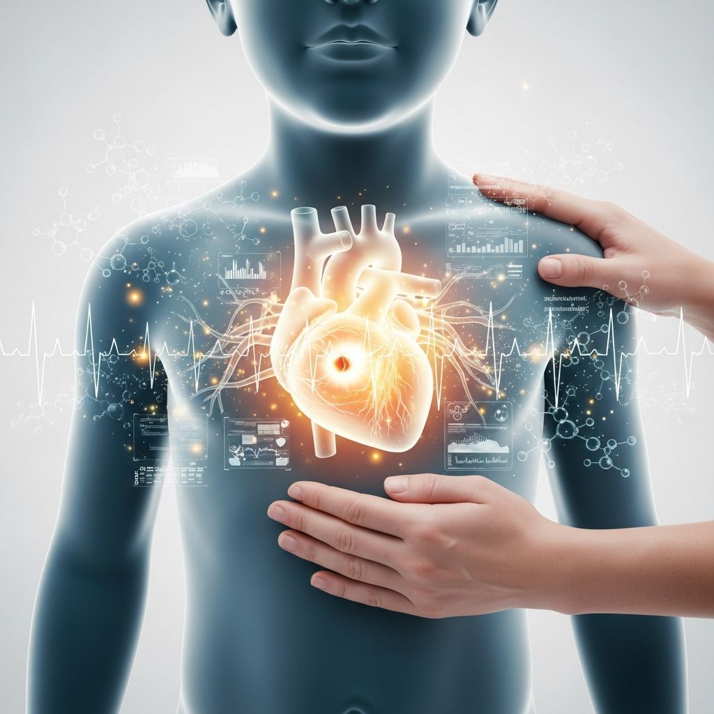
Congenital heart defects (CHDs) occur when a baby’s heart or major blood vessels do not form correctly during pregnancy, affecting about 1 in every 100 live births. These conditions vary widely in severity, from minor issues that may resolve without intervention to complex anomalies requiring multiple surgeries.
Understanding the Developing Heart
The human heart begins forming just weeks after conception, starting as a simple tube that folds and partitions into four chambers: two atria and two ventricles. Blood vessels and valves develop simultaneously to ensure efficient circulation of oxygen-rich blood to the body and oxygen-poor blood to the lungs. Disruptions during this critical window—often before a parent knows about the pregnancy—can lead to structural abnormalities.
Genetic factors play a role in many cases, including chromosomal changes like microdeletions seen in DiGeorge syndrome (22q11.2 deletion) or Williams syndrome (7p11.23 microdeletion), as well as single-gene mutations such as those in fibrillin-1 for Marfan syndrome or TBX5 for Holt-Oram syndrome. Environmental influences and unknown causes contribute to the rest.
Prevalence and Common Types
CHDs are the most frequent birth defects, with ventricular septal defects (VSDs)—holes between the heart’s lower chambers—being the most common, followed by atrial septal defects (ASDs). Cyanotic defects like tetralogy of Fallot occur less frequently but demand urgent care.
| Type | Description | Frequency |
|---|---|---|
| Ventricular Septal Defect (VSD) | Hole in the wall between ventricles, causing left-to-right shunting. | Most common in infancy |
| Atrial Septal Defect (ASD) | Hole between atria, often secundum type. | High prevalence |
| Tetralogy of Fallot | Four defects including pulmonary stenosis and VSD, causing cyanosis. | Most common cyanotic CHD |
| Transposition of Great Arteries | Aorta and pulmonary artery switched. | Less common but critical |
| Coarctation of Aorta | Narrowing of the aorta. | Obstructive lesion |
This table summarizes key examples; over 40 types exist, classified as cyanotic (causing blue skin from low oxygen) or acyanotic (obstructive or shunting).
Recognizing Symptoms in Newborns and Infants
Many CHDs show signs soon after birth. Cyanotic defects may present with bluish skin (cyanosis), especially during crying or feeding. Acyanotic ones often cause rapid breathing, poor weight gain, or fatigue during feeds due to heart strain.
- Cyanosis: Blue tint to lips, nails, or skin, indicating poor oxygenation.
- Breathing difficulties: Fast or labored breathing, grunting, or flaring nostrils.
- Feeding issues: Sweating, tiring quickly while nursing.
- Growth delays: Failure to thrive, pale appearance.
- Murmurs: Whooshing sounds heard via stethoscope during routine checks.
In obstructive lesions like aortic stenosis (3-6% of CHDs) or pulmonic stenosis (8-12%), murmurs arise from turbulent blood flow, potentially leading to heart failure if severe.
Diagnostic Approaches
Diagnosis often starts prenatally via ultrasound around 18-22 weeks, detecting many defects. Postnatally, tools include:
- Echocardiogram: Ultrasound imaging of heart structure and function.
- Chest X-ray: Checks heart size and lung fluid.
- ECG: Monitors electrical activity for arrhythmias.
- Cardiac catheterization: Invasive test for pressure measurements and imaging.
Early detection improves outcomes, with programs like those from Children’s Mercy emphasizing parent guides for prenatal and postnatal diagnosis.
Treatment Pathways: From Medications to Surgery
Treatment depends on defect severity. Simple ones like small VSDs or patent ductus arteriosus (PDA) may close spontaneously or with medication like indomethacin. Complex cases require surgery.
Medications: Prostaglandins keep ducts open in duct-dependent lesions; diuretics reduce fluid overload; ACE inhibitors ease heart workload.
Interventional procedures: Catheter-based fixes for stenoses or small shunts using balloons or devices.
Open-heart surgery: Repairs like patching VSDs or reconstructing vessels in tetralogy of Fallot. Critical neonates may need staged surgeries, as in hypoplastic left heart syndrome (HLHS).
Heart failure, a risk in volume-overload defects, manifests as pulmonary congestion or edema and requires prompt intervention.
Long-Term Management and Complications
Post-treatment, children need regular cardiology follow-ups. Risks include arrhythmias, pulmonary hypertension, endocarditis, and heart failure. Lifestyle advice includes antibiotic prophylaxis before dental work, activity restrictions if needed, and monitoring for iron overload from transfusions.
Support resources abound: American Heart Association for education, Mended Little Hearts for peer groups, and NHLBI for overviews on living with CHD.
Genetic and Syndromic Associations
Up to 30% of CHDs link to syndromes. Noonan syndrome (PTPN11 mutations) features pulmonic stenosis; Marfan involves aortic issues. Counseling helps families assess recurrence risks.
Emotional Support for Families
A CHD diagnosis overwhelms parents. Resources like Children’s Mercy’s Thrive Program offer guides on adjusting post-surgery and tube feeding, created with parent input. Connecting via support groups fosters resilience.
Frequently Asked Questions (FAQs)
What causes congenital heart defects?
Most result from complex genetic and environmental interactions during early fetal development, though many cases have no identifiable cause.
Can CHDs be prevented?
No guaranteed prevention, but folic acid intake before and during pregnancy may lower risks. Avoid teratogens like certain medications.
Will my child need lifelong medication?
Some do for blood pressure or clotting; others not after repair. Regular check-ups determine needs.
How do CHDs affect school and sports?
Many lead normal lives with modifications. Cardiologists guide activity levels to prevent strain.
What is the prognosis?
Over 90% survival to adulthood with modern care, though some need ongoing monitoring.
Advancing Care Through Research
Collaboratives like the National Pediatric Cardiology Quality Improvement Collaborative drive better outcomes via surveillance and family-centered care. NHLBI supports trials on long-term effects.
References
- Overview of Congenital Cardiovascular Anomalies — Merck Manual Professional Edition. 2023. https://www.merckmanuals.com/professional/pediatrics/congenital-cardiovascular-anomalies/overview-of-congenital-cardiovascular-anomalies
- About Congenital Heart Defects — American Heart Association. 2024-01-05. https://www.heart.org/en/health-topics/congenital-heart-defects/about-congenital-heart-defects
- Understanding Congenital Heart Defects — Children’s Mercy. 2023. https://www.childrensmercy.org/departments-and-clinics/heart-center/thrive-program/understanding-congenital-heart-defects/
- Congenital Heart Defects Infographic — CardioSmart.org (American College of Cardiology). 2022. https://www.cardiosmart.org/assets/infographic/congenital-heart-defects
- Congenital Heart Defects Resources — Centers for Disease Control and Prevention (CDC). 2025-06-12. https://www.cdc.gov/heart-defects/communication-resources/index.html
- Congenital Heart Disease — National Heart, Lung, and Blood Institute (NHLBI), NIH. 2024. https://www.nhlbi.nih.gov/health/congenital-heart-defects/types