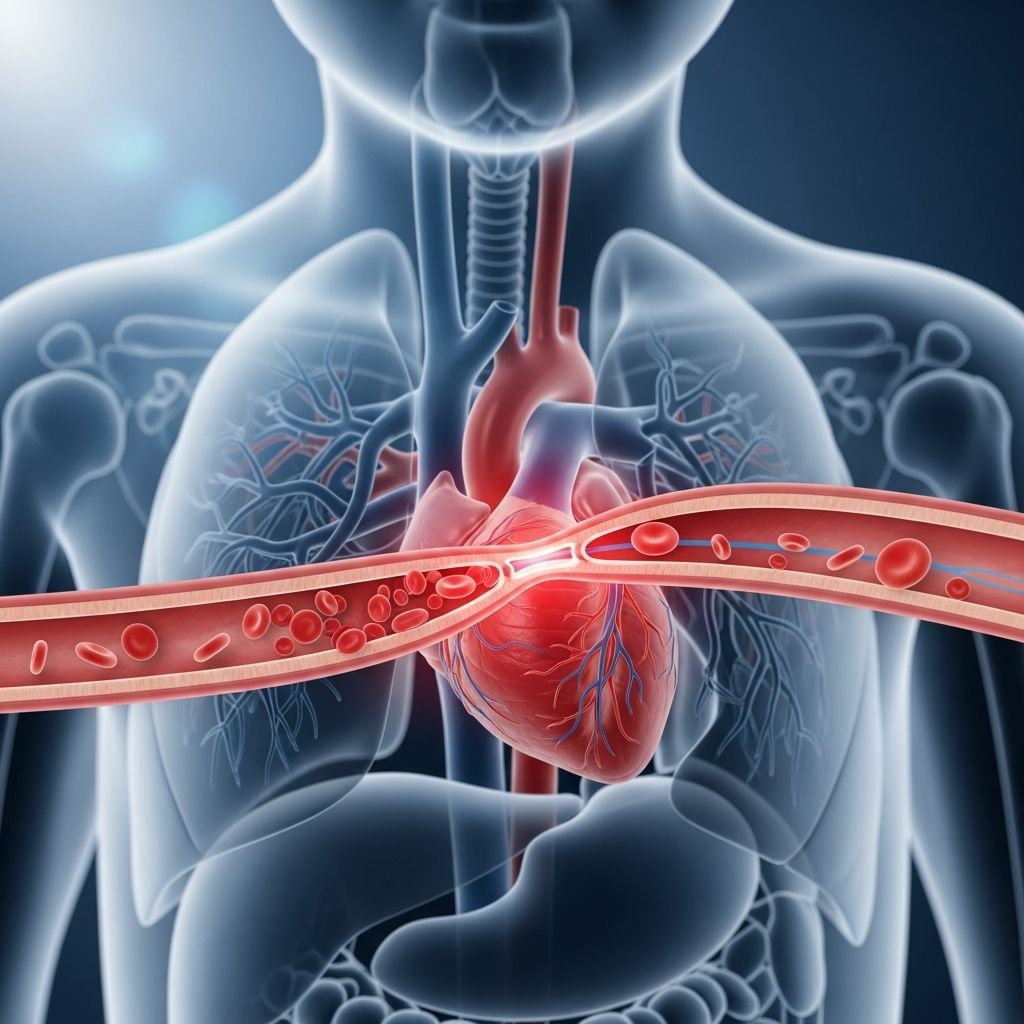
Coarctation of the aorta (COA) represents a common congenital heart anomaly where the body’s primary artery narrows, impeding blood flow. This condition affects approximately 1 in 1,700 newborns in the United States, often coexisting with other cardiac issues. Early detection and intervention are crucial to avert severe complications like heart failure or hypertension.
Understanding the Anatomy and Defect
The aorta serves as the central conduit for oxygen-rich blood pumped from the heart’s left ventricle to vital organs. In COA, a focal constriction typically arises near the ductus arteriosus insertion site, just beyond branches supplying the head and arms but proximal to those feeding the lower body. This bottleneck elevates pressure upstream (upper body) while starving downstream tissues (lower body), straining the heart over time.
Severity spans a spectrum: mild cases may evade notice until adolescence, whereas critical narrowings manifest in neonates as the fetal circulatory shunts close post-birth. Males predominate, comprising about two-thirds of cases, with familial patterns and associations to syndromes like Turner syndrome heightening risk.
Recognizing Warning Signs Across Age Groups
Symptoms hinge on constriction degree and age. Newborns with grave COA often crash into heart failure within days of birth, displaying:
- Rapid or labored breathing due to pulmonary congestion.
- Excessive sweating and irritability from metabolic distress.
- Poor appetite and faltering growth as energy diverts to the heart.
- Ashen or bluish skin tint signaling inadequate perfusion.
- Diminished or absent femoral pulses contrasting robust brachial ones.
Older children or teens with subtler narrowings might remain asymptomatic until routine checks uncover discrepancies. Common flags include:
- Leg cramps during activity (claudication) from muscle ischemia.
- Headaches, nosebleeds, or dizziness tied to hypertension.
- Chest discomfort on exertion.
- Cold extremities below the waist.
- Audible heart murmur during auscultation.
Undiagnosed adults face amplified risks, including aortic rupture or cerebral aneurysms, underscoring proactive screening.
Diagnostic Pathways for Timely Intervention
Clinicians initiate evaluation via comprehensive history and physical scrutiny, probing family cardiac history and growth metrics. Vital hallmarks are upper-lower limb blood pressure gradients exceeding 20 mmHg and weaker lower pulses—a “red flag” prompting specialist referral.
Noninvasive imaging anchors confirmation:
- Echocardiography: Gold standard, visualizing the narrowing, ventricular function, and collaterals via ultrasound.
- Chest X-ray: May reveal cardiomegaly or “rib notching” from enlarged intercostals in chronic cases.
- CT/MRI Angiography: Delineate anatomy preoperatively, especially complex variants.
- ECG: Detects left ventricular hypertrophy from pressure overload.
Prenatal echoes occasionally spot COA, while newborn pulse oximetry screens flag critical defects via desaturation.
Potential Health Risks if Left Unaddressed
| Complication | Description | Consequence |
|---|---|---|
| Hypertension | Chronic upper body high pressure | Heart strain, stroke risk |
| Heart Failure | Left ventricle overload | Poor pumping, fluid buildup |
| Renal Dysfunction | Low lower body perfusion | Kidney damage |
| Aortic Issues | Dilation, dissection | Rupture, death |
| Coronary Disease | Accelerated atherosclerosis | Heart attacks |
These perils escalate without repair, potentially culminating in fatality, particularly in infancy.
Treatment Options: From Catheter to Scalpel
Definitive management targets relief of obstruction, tailored to anatomy and patient stability. Medical stabilization precedes procedures in sick infants, employing prostaglandins to reopen the ductus and boost lower body flow alongside diuretics and inotropes.
Surgical Repair: Traditional mainstay, excising the coarcted segment and anastomosing healthy aorta ends. Success rates exceed 95%, with subclavian flap or patch aortoplasty variants for infants. Timing favors early correction to normalize hemodynamics.
Balloon Angioplasty: Catheter-delivered dilation suits recoarctations or select natives, though restenosis risks higher (10-20%). Stents enhance durability in adolescents.
Post-procedure, antihypertensive therapy and endocarditis prophylaxis guard against residuals.
Life After Correction: Monitoring and Lifestyle
Most repaired children thrive, engaging in sports sans restriction post-clearance. Yet, lifelong vigilance is imperative:
- Serial echoes track gradients and ventricular mass.
- Biennial blood pressure surveillance across limbs.
- Activity counseling, emphasizing hydration in heat.
- Subacute bacterial endocarditis precautions for dental work.
- Genetic consultation if syndromic features.
Hypertension recurs in 10-30%, demanding vigilant follow-up into adulthood.
FAQs for Worried Parents
Can COA resolve without surgery?
No, the structural narrowing persists, risking progressive damage despite collateral growth.
Is COA hereditary?
Not strictly, but familial clusters occur; Turner syndrome elevates odds.
What activities should my child avoid post-repair?
None typically, but consult cardiology before contact sports if residuals linger.
How do I check pulses at home?
Compare femoral (groin) to radial (wrist); asymmetry warrants medical review.
Does COA affect lifespan?
With timely repair and monitoring, prognosis excels; untreated cases falter.
Empowering Parents: Next Steps
Secure a pediatric cardiologist referral promptly upon suspicion. Multidisciplinary teams at congenital heart centers optimize outcomes. Educate on medications, warning signs like sudden breathlessness, and appointment adherence. Support groups foster resilience amid uncertainty.
In summary, COA, though daunting, yields to modern care. Informed vigilance transforms potential tragedy into routine success, letting kids flourish.
References
- Coarctation of the Aorta (COA) in Children — UR Medicine / University of Rochester Medical Center. 2023. https://www.urmc.rochester.edu/encyclopedia/content?contenttypeid=90&contentid=P01770
- Coarctation of the Aorta (COA) — Arkansas Children’s Hospital. 2024. https://www.archildrens.org/programs-and-services/cardiology/Conditions/coarctation-of-the-aorta
- Coarctation of the Aorta (CoA) — Cleveland Clinic. 2024-01-15. https://my.clevelandclinic.org/health/diseases/16876-aortic-coarctation
- Coarctation of the Aorta — Nemours KidsHealth. 2023. https://kidshealth.org/en/parents/coa.html
- Coarctation of the Aorta (CoA) — American Heart Association. 2024. https://www.heart.org/en/health-topics/congenital-heart-defects/about-congenital-heart-defects/coarctation-of-the-aorta-coa