What Parents Should Know About Bladder Ultrasound
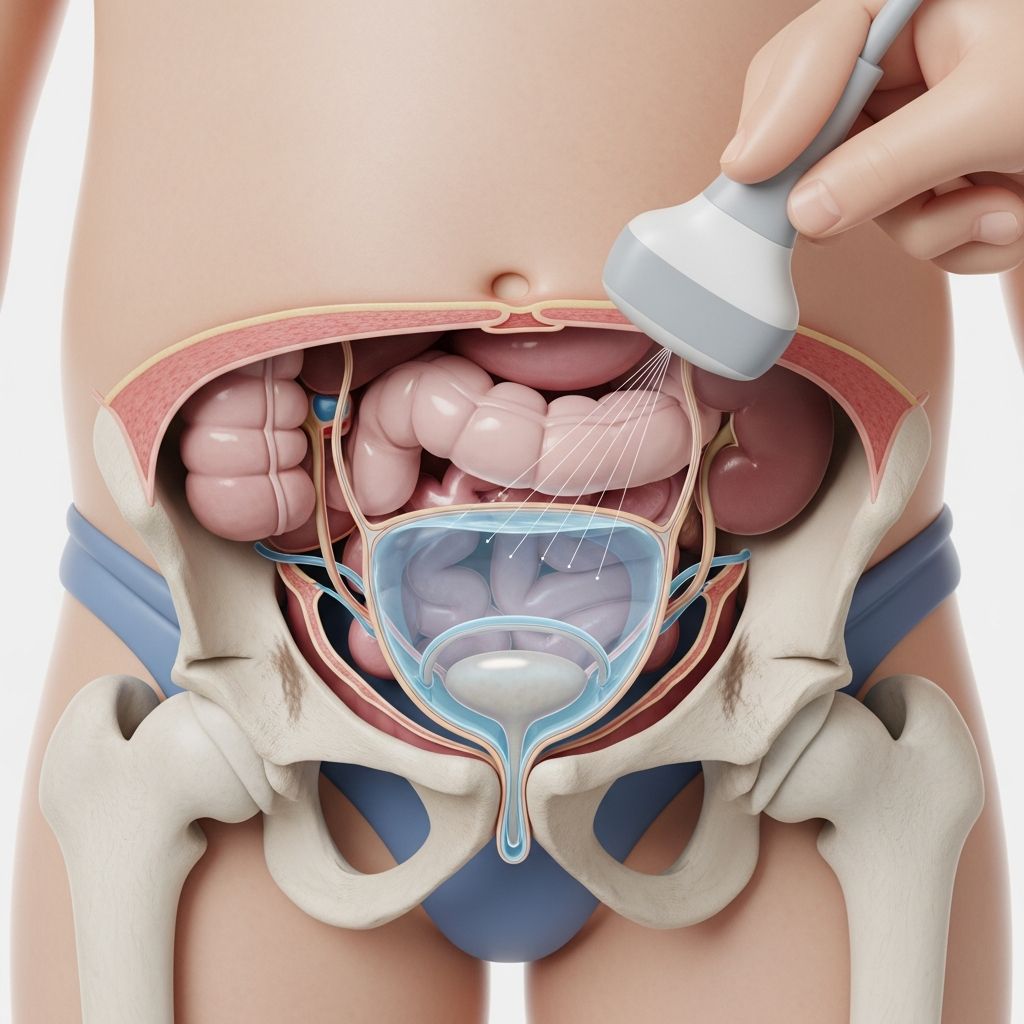
A bladder ultrasound is a medical imaging technique that uses sound waves to create pictures of your child’s bladder and the surrounding urinary structures. Unlike X-rays or CT scans, this imaging method does not expose your child to radiation, making it a safe and comfortable option for evaluating urinary concerns. The procedure creates visual images by transmitting high-frequency sound waves through a handheld device called a transducer, which bounces off the bladder and surrounding tissues to produce detailed images on a monitor.
This non-invasive examination helps doctors understand how your child’s bladder is functioning and can identify various conditions that might be affecting urinary health. Parents often feel anxious about medical procedures, but understanding what to expect can help ease concerns and prepare your child for the appointment.
Why Doctors Recommend Bladder Ultrasound for Children
Healthcare providers may recommend a bladder ultrasound when a child experiences specific urinary symptoms or when they need to monitor known bladder conditions. Common reasons for ordering this imaging study include:
- Persistent bedwetting or daytime accidents: When children continue to have involuntary urination beyond the typical age of bladder control, investigating the bladder’s structure and function becomes important.
- Urinary tract infections: Recurrent or severe infections may indicate underlying anatomical problems that need visualization.
- Difficulty emptying the bladder: Some children struggle to completely empty their bladder, which can be evaluated through ultrasound measurements.
- Painful urination: When a child experiences pain during the act of urinating, imaging helps rule out physical causes.
- Blood in the urine: The presence of blood requires investigation to determine the source and cause.
- Abdominal or pelvic discomfort: Unexplained pain in these areas may be related to bladder function or structure.
- Monitoring after treatment: Children who have previously been diagnosed with bladder conditions may need follow-up imaging to ensure healing and proper function.
- Evaluating congenital conditions: Some children are born with structural variations of the urinary tract that require ongoing assessment.
Preparing Your Child for the Procedure
Proper preparation is essential for obtaining clear ultrasound images and ensuring your child feels comfortable during the examination. The most important preparation step involves managing your child’s bladder fullness.
Getting Your Child Ready at Home
Before leaving for the appointment, your child should drink plenty of water or clear fluids several hours before the scheduled ultrasound. The specific amount depends on your child’s age, and your healthcare provider will give you detailed instructions. A full bladder improves image quality because sound waves travel more effectively through liquid, providing clearer pictures for the radiologist to analyze.
Help your child understand that they should not empty their bladder before arriving at the imaging center. This might feel uncomfortable, but it is necessary for the procedure’s success. Explaining this in age-appropriate language can help your child cooperate. You might say something like, “The doctor needs to see your bladder when it is full, so we want to make sure you have plenty of liquid in there.”
What to Wear and Bring
Dress your child in comfortable, easy-to-remove clothing that allows access to the lower abdomen and pelvic area. Avoid clothing with complicated fasteners or multiple layers. Bring your child’s insurance card and any identification documents required by the imaging facility. Some centers request that you arrive 10-15 minutes early to complete paperwork.
What Happens During the Bladder Ultrasound
Understanding the step-by-step process can help you prepare your child mentally and emotionally for the appointment.
The Initial Steps
When you arrive at the ultrasound department, a trained sonographer will greet you and your child. They will explain the procedure in simple terms appropriate for your child’s age. Some imaging centers allow parents to remain in the room during the scan, which can provide comfort and reassurance to anxious children.
Your child will be asked to lie on a comfortable examination table on their back. The sonographer may ask your child to lift their shirt to expose the lower abdomen and pelvic area. A clean, water-based gel will be applied to the skin over the lower belly. This gel feels cool initially but warms quickly. Explain to your child beforehand that they will feel the gel and the gentle pressure of the transducer moving across their skin, but the procedure should not cause pain.
The Scanning Process
The sonographer will move the transducer slowly and deliberately across the lower abdomen, starting near the belly button and moving toward the pubic bone. They will apply gentle pressure while angling the transducer to capture images from different perspectives. Your child may see images appearing on the monitor during the scan. Many children find it interesting and even educational to watch their own bladder appear on the screen.
The procedure typically takes 15-30 minutes, depending on how quickly clear images can be obtained and whether additional measurements are needed. Your child will hear a soft whooshing or swishing sound as the ultrasound machine captures the images. This is completely normal and nothing to worry about.
Post-Void Measurement
In many cases, the sonographer will perform an initial measurement of the bladder while it is full. Then, your child will be directed to a restroom to empty their bladder completely. After your child returns, a second measurement is taken to assess how much urine remains. This measurement, called post-void residual volume, helps doctors understand how effectively your child’s bladder is emptying.
Understanding the Results
After the scan is complete, the sonographer will wipe away the gel, and your child can get dressed. The images will be reviewed by a radiologist, a physician who specializes in interpreting medical imaging. Results are typically communicated to your child’s primary care physician or the specialist who ordered the test.
Normal Findings
When results are normal, the radiologist’s report may use terms such as “no acute findings,” “normal findings,” “unremarkable,” or “no abnormality detected.” These terms indicate that the bladder appears structurally normal, the bladder wall thickness is appropriate, the bladder capacity is typical for your child’s age, and the post-void residual volume is within expected ranges.
Abnormal Findings and What They Mean
If the ultrasound reveals abnormal findings, your healthcare provider will discuss the results with you and explain what they mean for your child’s health. Possible abnormal findings include:
- Bladder diverticula: Small pouches or outpouchings in the bladder wall that may trap urine and contribute to infections.
- Excessive post-void residual volume: When too much urine remains after emptying, which could indicate obstruction, nerve problems, or weak bladder muscle function.
- Structural abnormalities: Variations present from birth, such as duplication of urinary structures or other congenital differences.
- Bladder wall thickening: May suggest chronic irritation or obstruction.
- Stones or deposits: Mineral accumulations that can form when urine becomes too concentrated.
- Masses or lesions: Unusual growths that require further evaluation.
When Additional Testing Becomes Necessary
Sometimes a bladder ultrasound provides initial information, but your child’s doctor may recommend additional testing to fully understand the condition.
Complementary Imaging Studies
A kidney ultrasound often accompanies bladder ultrasound because kidney and bladder problems frequently occur together. While a bladder ultrasound focuses primarily on the bladder itself, a kidney ultrasound examines the kidneys and the connecting tubes called ureters. Many imaging centers perform both studies during a single appointment by repositioning the transducer over the kidney area.
CT scans provide three-dimensional images using X-ray technology and offer greater anatomical detail than ultrasound. These are typically ordered when ultrasound findings are unclear or when more precise measurements are needed before surgical intervention.
MRI scans use magnetic and radio waves to create detailed internal images without radiation exposure. These are particularly useful for evaluating soft tissues and can provide information that ultrasound cannot capture.
Functional and Laboratory Tests
To assess how the bladder actually functions during the act of urination, doctors may recommend urodynamic testing. This specialized test measures bladder pressure and flow rates during voiding.
A urinalysis examines a urine sample under a microscope and through chemical testing to identify signs of infection, blood, crystals, or other abnormalities.
Cystoscopy involves inserting a thin, lighted tube into the urethra to directly visualize the inside of the bladder. This procedure allows the physician to see structures that ultrasound cannot and to take tissue samples if needed.
Helping Your Child Feel Comfortable
Medical procedures can cause anxiety in children. Several strategies can help ease your child’s experience:
- Prepare your child in advance by explaining what will happen using simple, non-threatening language.
- Practice the positioning at home so your child knows what to expect.
- Bring a comfort item such as a favorite stuffed animal or blanket if the facility permits.
- Use positive reinforcement and praise your child for cooperating.
- Ask the sonographer if your child can listen to music or watch a show during the procedure.
- Remain calm yourself, as children often pick up on parental anxiety.
- Arrange a small reward after the appointment to give your child something to look forward to.
After the Procedure
Once the ultrasound is complete, your child can return to normal activities immediately. There are no restrictions on eating, drinking, or physical activity. The gel used during the procedure washes off easily with soap and water. Some children feel slightly uncomfortable from the full bladder during the scan, but this sensation disappears once they empty their bladder.
Your healthcare provider will contact you with results within a few days. If your child’s doctor needs to discuss the findings or recommend treatment, they will schedule a follow-up appointment.
Frequently Asked Questions
Is bladder ultrasound safe for children?A: Yes, bladder ultrasound is very safe. It uses sound waves rather than radiation, making it an excellent choice for pediatric imaging. The procedure is painless and non-invasive.
Q: How long does a bladder ultrasound take?
The procedure typically takes 15-30 minutes, depending on how quickly clear images are obtained and whether additional measurements are needed.
Will my child feel pain during the ultrasound?
No, the procedure should not cause pain. Your child may feel the cool gel and gentle pressure from the transducer, but these sensations are not painful. The full bladder may feel uncomfortable, but this resolves once emptying occurs.
What if my child has difficulty drinking enough fluids before the appointment?
Contact the imaging center to discuss your concerns. They may provide alternative preparation instructions or allow extra time for your child to drink fluids at the facility.
Can I stay in the room during the ultrasound?
Most imaging centers allow parents to remain in the room to provide reassurance. Ask about this policy when scheduling your appointment.
How soon will I get the results?
Results are typically available within a few days. The radiologist’s report will be sent to your child’s healthcare provider, who will discuss the findings with you.
Are there any risks associated with bladder ultrasound?
Bladder ultrasound carries virtually no risks. There is no radiation exposure, no need for injections or catheters, and no known adverse effects from sound waves.
References
- Bladder Ultrasound: Procedure, Preparation and Results — PocketHealth. 2025. https://www.pockethealth.com/patient-resources/bladder-ultrasound-how-to-prepare-and-what-to-expect/
- Bladder Ultrasound: Purpose, Procedure, Results & More — Healthline. 2024. https://www.healthline.com/health/bladder-ultrasound
- Bladder Ultrasound Made Easy: Step-By-Step Guide — POCUS 101. 2025. https://www.pocus101.com/bladder-ultrasound-made-easy-step-by-step-guide/
- What To Expect: Bladder Ultrasound — NYHealth.com. 2024. https://nyhealth.com/what-to-expect/bladder-ultrasound
- Portable Bladder Ultrasound: An Evidence-Based Analysis — National Center for Biotechnology Information. 2012. https://pmc.ncbi.nlm.nih.gov/articles/PMC3379524/
- Renal (Kidney) Ultrasound — Radiologyinfo.org. 2024. https://www.radiologyinfo.org/en/info/ultrasound-renal