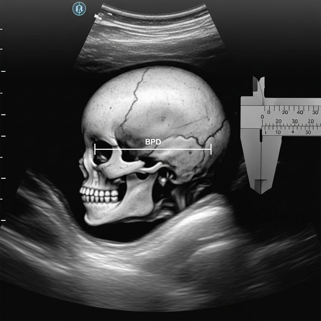
The biparietal diameter (BPD) is a fundamental ultrasound measurement that gauges the width of a fetus’s head, playing a central role in prenatal care by helping estimate gestational age and monitor growth patterns. This metric, captured between the parietal bones of the skull, offers healthcare providers critical data on fetal development throughout pregnancy.
Understanding Fetal Biometry Fundamentals
Fetal biometry involves precise ultrasound assessments of various body parts to evaluate size, proportions, and growth velocity. These scans, typically starting around 13 weeks, compare measurements against standardized charts to assign percentiles and detect deviations early. BPD stands out as one of the core parameters, alongside others that provide a holistic view of fetal health.
During routine prenatal visits, sonographers use high-frequency sound waves to visualize the fetus in a transverse plane, ensuring the head appears as a perfect circle for accurate BPD capture. This non-invasive process allows real-time analysis, often combined with additional metrics for comprehensive insights.
Defining Biparietal Diameter in Detail
BPD specifically measures the transverse distance across the fetal skull at its widest point, from the outer edge of one parietal bone to the other. Known as the ‘outer-to-outer’ method (BPDoo), this approach ensures consistency and reproducibility across scans.
This measurement proves invaluable for several reasons. It aids in confirming gestational age, particularly between 11 and 16 weeks when head growth follows a predictable trajectory. Moreover, BPD contributes to fetal weight estimation formulas, integrating with other biometrics for reliable predictions.
- Gestational dating: BPD helps refine due dates when combined with early pregnancy data.
- Growth tracking: Serial measurements reveal if the fetus is growing appropriately.
- Anomaly detection: Abnormal sizes may signal conditions like microcephaly (small head) or macrocephaly (large head).
Standard BPD Values Across Pregnancy Stages
BPD values evolve predictably with gestation. In the second trimester, for instance, a 20-week fetus typically shows a BPD around 46-50 mm, increasing steadily to about 90-95 mm by 40 weeks. These ranges derive from population-based charts, adjusted for factors like maternal ethnicity and equipment calibration.
| Gestational Age (Weeks) | Mean BPD (mm) | 5th-95th Percentile (mm) |
|---|---|---|
| 16 | 34 | 29-39 |
| 20 | 47 | 42-52 |
| 24 | 59 | 53-65 |
| 28 | 70 | 63-77 |
| 32 | 79 | 72-86 |
| 36 | 87 | 79-95 |
| 40 | 94 | 86-102 |
These figures represent averages from clinical guidelines; individual variations occur, but values outside the 5th-95th percentiles warrant further evaluation.
Complementary Measurements in Fetal Assessment
No single metric tells the full story—BPD works in tandem with others for balanced analysis. Head circumference (HC) traces the outer skull perimeter, offering a complementary view of brain growth. Femur length (FL) assesses long bone development, while abdominal circumference (AC) reflects nutritional status and organ size.
- HC: Measures full head oval, useful for detecting shape irregularities.
- FL: Indicates skeletal maturation, correlating with height potential.
- AC: Largest single predictor of fetal weight, sensitive to growth restriction.
Ratios like cephalic index (BPD to occipitofrontal diameter) further refine head shape evaluations, ensuring proportional development.
Performing the BPD Ultrasound Procedure
Ultrasound for BPD requires optimal fetal positioning. The mother lies supine as gel is applied to the abdomen, and a transducer glides to capture a clear coronal view of the thalami and cavum septi pellucidi. Callipers are placed precisely on the leading edges of the parietal bones.
Transvaginal approaches suit early pregnancies (under 14 weeks), transitioning to abdominal scans later. Modern machines auto-calculate derived values like HC from BPD and occipitofrontal diameter (OFD), streamlining workflows.
Expect 20-45 minutes per session, with multiple images archived for serial comparison. Patient comfort is prioritized, minimizing movement for crisp images.
Interpreting BPD Results and Clinical Implications
Results are plotted on growth charts, assigning percentiles. A 50th percentile BPD signifies average growth; extremes prompt investigations like Doppler studies or amniocentesis. For example, lagging BPD might indicate intrauterine growth restriction (IUGR), while accelerated growth could suggest macrosomia.
Fetal weight estimates (EFW) incorporate BPD via formulas like Hadlock 4: EFW = 10^ (1.3596 – 0.00386 AC FL + 0.0064 HC + 0.00061 BPD AC + 0.0424 AC + 0.174 FL). Consistency across methods validates predictions, with BPDoo preferred for interoperability.
Timing and Frequency of Biometry Scans
Early scans (6-14 weeks) focus on crown-rump length (CRL) for dating, with BPD reliable post-13 weeks. Anatomy scans at 18-22 weeks introduce full biometry, followed by growth checks at 28, 32, and 36 weeks in high-risk cases.
Low-risk pregnancies may suffice with two ultrasounds, but conditions like diabetes or hypertension necessitate more frequent monitoring to track BPD trends.
Potential Limitations and Accuracy Factors
BPD accuracy hinges on fetal lie, operator skill, and equipment quality. Intra- and inter-observer variability is low (around 1-2 mm) with standardized protocols, but ossification delays or dolichocephaly can skew readings.
Post-26 weeks, FL and AC gain prominence as BPD growth plateaus. Always contextualize with multiple parameters to avoid misdiagnosis.
Advanced Applications of BPD Data
Beyond basics, BPD informs research on ethnic-specific growth curves and AI-enhanced predictions. Integration with 3D ultrasound refines measurements, while serial BPD tracks interventions for growth disorders.
In preterm labor assessments, BPD aids viability estimates, guiding neonatal care planning.
Frequently Asked Questions About BPD in Pregnancy
What does a small BPD indicate?
A BPD below the 10th percentile may suggest microcephaly, IUGR, or dating errors; further tests clarify causes.
Can BPD alone determine fetal weight?
No, it combines with HC, AC, and FL for accurate EFW via validated equations.
Is BPD measured in every ultrasound?
Typically from the second trimester onward in standard biometry scans.
How reliable is BPD for due date prediction?
Highly reliable mid-trimester, less so later; early CRL supersedes it for first-trimester dating.
Does BPD predict delivery complications?
Large BPD correlates with cephalopelvic disproportion risks, prompting cesarean discussions.
Empowering Parents with Ultrasound Insights
Understanding BPD equips expectant parents to engage meaningfully with providers. Request percentile charts and trend graphs during appointments to visualize progress. Digital platforms like patient portals facilitate result access, fostering informed decisions.
While empowering, remember measurements guide—not dictate—care. Consult obstetricians for personalized interpretations amid normal variations.
References
- Fetal Biometry: What is BPD on an Ultrasound? — PocketHealth. 2023. https://www.pockethealth.com/patient-resources/fetal-ultrasound/
- What is BPD in Pregnancy? — Mamata Fertility Hospital. 2024. https://mamatafertility.com/blog/what-is-bpd-in-pregnancy/
- Biparietal diameter measurements using the outer‐to‐outer versus inner‐to‐outer calliper placement method. — PMC (Wiley). 2021-09-14. https://pmc.ncbi.nlm.nih.gov/articles/PMC8409806/
- BPD Measurements. — Clarius Mobile Health. 2023. https://clarius.com/classroom/bpd-measurements/
- Fetal Biometry: Measurements, Normal Values & Accuracy. — Cleveland Clinic. 2023-07-20. https://my.clevelandclinic.org/health/diagnostics/25087-fetal-biometry
- Fetal size and dating: charts recommended for clinical obstetric practice. — BMUS. 2009-08. https://www.bmus.org/static/uploads/resources/Aug_2009_Fetal_Measurements_D3NApK5.pdf