Arteriovenous malformations, often shortened to AVMs, are unusual tangles of blood vessels that can change how blood flows through a child’s body. They can sound frightening, but learning the basics helps parents stay calm, ask good questions, and partner closely with doctors.
This guide explains AVMs in clear, family-friendly language. It focuses on how AVMs show up in children, how they are found, treatment choices, and what daily life can look like before and after treatment.
How Blood Vessels Normally Work
To understand AVMs, it helps to picture how healthy blood vessels form a loop. Arteries carry blood away from the heart under higher pressure, a tiny network of capillaries delivers oxygen and nutrients to tissues, and veins bring blood back to the heart at lower pressure.
- Arteries: Thick, muscular tubes that handle strong pulses of blood from the heart.
- Capillaries: Very small vessels where the real “exchange” of oxygen, nutrients, and waste happens with nearby cells.
- Veins: Softer vessels that return blood to the heart after the tissues have used what they need.
The capillary network is important because it slows blood down and spreads it out. This protects delicate tissues from high pressure and makes sure they get enough oxygen.
What Is an Arteriovenous Malformation?
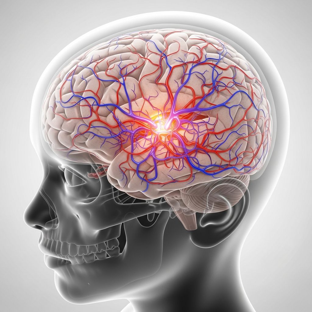
An arteriovenous malformation is a cluster of arteries and veins that are directly connected to each other without the usual capillaries in between. Instead of blood flowing through a gentle, fine web of tiny vessels, it rushes straight from artery to vein.
This shortcut can cause several problems:
- Blood moves too quickly, giving nearby tissues less time to receive oxygen and nutrients.
- The high-pressure flow can stretch and weaken vessel walls over time.
- The heart may need to work harder to keep up, especially with very large AVMs.
AVMs can appear almost anywhere blood vessels exist, including the skin, muscles, organs, brain, or spinal cord. In children, they are usually something a child is born with, even if they are not noticed right away.
Are AVMs the Same as Birthmarks?
Some AVMs sit close to the skin and may first look like a birthmark or patch of colored skin. However, not all birthmarks are AVMs, and not all AVMs change skin color. A careful exam and imaging are needed to tell them apart from other vascular marks.
Common visible clues of a possible AVM near the skin can include:
- A pink, red, or purple area that seems deeper or more “pulsing” than a typical flat birthmark.
- Skin that feels warmer or thicker than the surrounding area.
- Swelling or a soft mass that may slowly grow over months or years.
Why AVMs Can Be a Concern
Many AVMs stay quiet for years and cause few problems. Others may lead to symptoms because of their location, size, or the strain they place on surrounding tissues and blood vessels.
Possible complications include:
- Bleeding: Fragile vessels can leak or break, especially in the brain or digestive tract.
- Pain and swelling: The rapid blood flow and extra blood volume can stretch tissues and cause discomfort.
- Effects on growth or function: Large AVMs in limbs or organs can change normal development or movement.
- Heart strain: Very large AVMs may make the heart pump harder, sometimes leading to heart problems if untreated.
Common Symptoms Parents May Notice
Symptoms depend mainly on where the AVM is located and how large or active it is. Some children show signs early in life, while others are not diagnosed until later childhood or adolescence.
General signs around the skin or limbs
- A patch of skin that looks red, purple, or darker than the surrounding area.
- Swelling or fullness that may feel soft or slightly bumpy.
- Warmth when touching the area compared to other parts of the body.
- A gentle vibration or pulse that can sometimes be felt with fingertips.
- Pain, especially with activity, growth spurts, or after minor bumps.
Possible signs of an AVM in the brain or spine
When AVMs form in the nervous system, they may disturb how nerves send and receive signals. Symptoms can vary widely, but may include:
- Frequent or worsening headaches.
- Seizures in a child with no prior seizure history.
- Weakness, numbness, or clumsiness in an arm or leg.
- Difficulty with balance, speaking, or understanding language.
- Changes in vision, such as blurred areas or missing parts of the visual field.
- Sudden vomiting, severe headache, or loss of consciousness (possible bleeding emergency).
When to seek urgent care
Call emergency services or go to the nearest emergency department if your child has:
- A sudden, extremely severe headache or back pain.
- A seizure lasting more than a few minutes or repeated seizures.
- Sudden weakness, drooping on one side of the face, or trouble speaking.
- Heavy bleeding from an area known or suspected to be an AVM.
How Doctors Diagnose AVMs
If a pediatrician suspects an AVM, your child will usually be referred to specialists such as pediatric neurologists, neurosurgeons, interventional radiologists, or vascular surgeons. Diagnosis often combines a physical exam with several imaging tests.
| Test | What it Shows | Typical Use |
|---|---|---|
| Ultrasound (Doppler) | Blood flow speed and direction in surface vessels | Initial look at AVMs in limbs or close to the skin |
| MRI or MRA | Detailed images of soft tissue and blood vessels | Brain, spine, deep soft tissues; planning treatment |
| CT or CTA scan | Cross-sectional pictures, sometimes with contrast dye | Quick overview in emergencies or complex cases |
| Catheter angiography | Real-time dye images of arteries and veins | Detailed map before procedures like embolization or surgery |
These tests help the team see how large the AVM is, which blood vessels are involved, whether there are signs of bleeding, and which treatment options may be safest and most effective.
Treatment Goals for AVMs in Children
The main goal of treatment is to lower the risk of bleeding and other long-term problems while protecting normal brain and body function. Because every child and every AVM is different, treatment plans are customized.
Depending on the situation, doctors may recommend:
- Watching the AVM over time with regular checkups.
- Blocking abnormal blood flow from the inside using special materials.
- Carefully removing the AVM with surgery.
- Using precisely targeted radiation to slowly close off the abnormal vessels.
Common Treatment Options Explained
1. Careful monitoring (watchful waiting)
Some small, stable AVMs that are hard to reach or are not causing symptoms may be safely monitored instead of treated right away. This approach helps avoid risks from procedures while still keeping a close eye on the AVM.
Monitoring can include:
- Regular checkups with a specialist.
- Repeat imaging tests every so often.
- Tracking any new pain, swelling, headaches, or changes in movement or behavior.
2. Endovascular embolization
In this minimally invasive procedure, a specialist threads a thin tube (catheter) through blood vessels, usually starting from the groin, up to the AVM. Tiny plugs, coils, or glue-like materials are then placed inside the abnormal vessels to reduce or stop the fast blood flow.
Key points about embolization:
- Often done under general anesthesia in a hospital.
- May be used on its own for some AVMs or as preparation before surgery or radiation.
- Can be repeated in stages for very complex or large AVMs.
3. Surgery
In some cases, surgeons can remove the AVM through an operation. This involves opening the skin (and sometimes the skull for brain AVMs), carefully separating the abnormal vessels from healthy tissue, and closing off the feeding arteries and draining veins.
Surgery may be considered when:
- The AVM is reachable without major damage to important brain or body structures.
- Bleeding risk is high or the AVM has already caused a serious hemorrhage.
- Other treatments alone are not enough to make the AVM safe.
4. Stereotactic radiosurgery (targeted radiation)
Stereotactic radiosurgery is not surgery in the usual sense. It uses tightly focused beams of radiation to damage the AVM from within. Over months to a few years, the treated vessels may thicken and close off, reducing risk of bleeding.
Highlights of this option:
- Often used for small to medium AVMs located deep in the brain.
- Done as an outpatient or short hospital stay in many centers.
- Bleeding risk does not disappear immediately; follow-up imaging is crucial.
Life at Home Before and After Treatment
Living with an AVM can feel stressful, especially right after diagnosis. Parents often worry about everyday activities, school, and sports. The right plan depends on the AVM’s size and location, and on the advice of the medical team.
Everyday lifestyle tips
- Follow all medication instructions, especially for seizure control or pain relief.
- Ask about safe levels of physical activity; gentle exercise is often encouraged.
- Help your child avoid head or limb injuries whenever possible (helmets, pads, safe play).
- Teach your child in age-appropriate ways about their condition so they can speak up if they feel unwell.
Emotional support for families
Children may feel anxious, different from their friends, or worried about tests and procedures. Parents can support them by listening, giving honest but reassuring information, and working with school counselors or psychologists when needed.
- Consider connecting with other families through reputable support groups.
- Ask the care team about child life specialists who can prepare kids for procedures using play and simple language.
Questions to Ask Your Child’s Care Team
Bringing written questions to visits can make appointments more productive and reduce stress. Useful topics include:
- Where exactly is my child’s AVM located, and how large is it?
- What symptoms should prompt an urgent call or emergency visit?
- What are the short- and long-term risks if we treat now versus monitor?
- Which treatments are being recommended and why?
- How many similar cases has this hospital or team treated?
- What should teachers and school nurses know about my child’s condition?
Long-Term Outlook
Advances in imaging, minimally invasive procedures, and neurosurgery have greatly improved outcomes for many children with AVMs. Some AVMs can be cured or greatly reduced in risk with modern treatments. Others can be managed safely for many years with careful follow-up.
Because a growing child’s body changes over time, long-term care usually includes periodic visits, imaging when indicated, and ongoing attention to learning, behavior, and development. Early support services, such as physical, occupational, or speech therapy, can help children reach their full potential.
Frequently Asked Questions About AVMs in Children
1. Did my child’s AVM come from something I did during pregnancy?
Current evidence suggests that most AVMs develop very early in pregnancy as blood vessels are forming. They are usually not linked to anything a parent did or did not do. For most families, there is no identifiable cause or way that the AVM could have been prevented.
2. Can AVMs be inherited?
Most AVMs occur in children with no family history of vascular problems. However, a few rare genetic conditions can increase the chance of developing abnormal blood vessels. If your child has multiple AVMs or other unusual findings, the medical team may suggest genetic counseling or testing.
3. Will my child always have restrictions on sports and play?
Some children with AVMs can join many typical activities, while others need to avoid certain contact sports or high-impact events. The recommendations depend on the AVM’s location, size, and the treatments planned or completed. Your child’s specialists are the best guides on which activities are safe and which should be limited.
4. Is surgery or treatment always needed?
No. In some cases, the safest option is to monitor the AVM over time instead of treating it right away. Doctors weigh the AVM’s chance of causing bleeding or other problems against the risks of each procedure. Parents are usually closely involved in choosing the approach that fits their child best.
5. Can an AVM grow or change as my child grows?
Some AVMs stay almost the same over time; others may slowly expand or change as a child grows and blood flow patterns shift. Regular follow-up visits and imaging, when recommended, help catch changes early so the plan of care can be adjusted if needed.
6. What should I tell my child’s school?
Most schools appreciate clear, simple information about any health condition that might affect safety or learning. Ask your healthcare team for a written summary explaining what an AVM is, which symptoms need quick attention, and any activity limits. Sharing this with teachers, coaches, and the school nurse helps create a safer, more supportive environment.
By staying informed, asking questions, and building a strong partnership with your child’s healthcare team, you can help your child live as safely and fully as possible with an AVM.