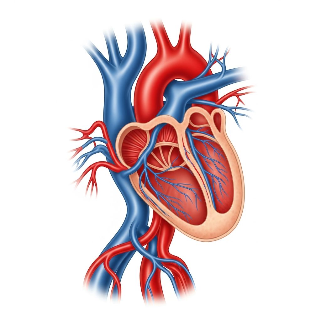
The aortic arch represents a critical curved segment of the body’s primary artery, the aorta, responsible for channeling oxygen-rich blood from the heart to the upper body regions in children. Positioned in the chest, it ensures vital circulation to the brain, arms, and neck, playing a foundational role in a child’s growth and daily activities.
Understanding the Aorta’s Path in Young Bodies
In pediatric anatomy, the aorta emerges from the left ventricle, initially ascending before forming the distinctive arch that bends leftward and backward. This structure transitions into the descending aorta, distributing blood efficiently throughout the torso and lower extremities. For children, maintaining this pathway’s integrity is essential for supporting rapid development and physical demands.
The arch’s positioning behind the breastbone and alongside the windpipe underscores its protected yet pivotal location within the chest cavity. Its layered composition—inner endothelial lining, muscular middle layer with elastic properties, and outer supportive tissue—allows it to withstand high-pressure blood flow while adapting to a growing child’s needs.
Key Branches Supplying Upper Body Needs
- Brachiocephalic Trunk: The initial and largest branch splits into the right subclavian artery (for the right arm) and right common carotid artery (for the right brain side and neck), ensuring robust supply to these areas.
- Left Common Carotid Artery: Arises next, delivering blood directly to the left brain hemisphere and neck structures, vital for cognitive and sensory functions in kids.
- Left Subclavian Artery: The final branch nourishes the left arm, supporting motor skills and play activities essential for childhood.
These branches emerge sequentially from the arch’s upper curve, optimizing blood delivery to regions above the heart level. Variations occur in about 20-30% of cases, where the left carotid might stem from a common trunk, but these rarely impact function in healthy children.
How the Aortic Arch Forms During Fetal Growth
Embryonic development involves a series of aortic arches derived from branchial arches, with the mature left fourth arch persisting as the definitive structure connecting ascending and descending portions. In utero, the ductus arteriosus—a temporary vessel—links the pulmonary artery to the arch’s base, shunting blood away from immature lungs.
Post-birth, this ductus typically closes within days, transforming into the ligamentum arteriosum, a fibrous remnant anchoring the arch. Disruptions in this process can lead to persistent patency, affecting neonatal circulation and requiring monitoring in infants.
Common Abnormalities Affecting Children’s Aortic Arch
Several congenital issues can alter the arch’s shape or function, often detected via prenatal or newborn screenings. These conditions may narrow blood flow, strain the heart, or cause pressure imbalances.
| Condition | Description | Impact on Kids |
|---|---|---|
| Coarctation of the Aorta | Narrowing typically at the arch’s lower curve, near ductus insertion site. | High blood pressure in arms, weak leg pulses; may cause heart strain or failure if severe. |
| Double Aortic Arch | Two arches encircling trachea/esophagus, forming vascular ring. | Breathing difficulties, swallowing problems, frequent respiratory infections in infants. |
| Right Aortic Arch | Arch curves rightward instead of left, sometimes with aberrant vessels. | Often asymptomatic but can compress nearby structures, leading to feeding issues. |
| Interrupted Aortic Arch | Complete gap in arch continuity, rare and critical. | Requires immediate surgery; presents with shock and poor perfusion. |
These anomalies arise from faulty embryonic remodeling, affecting roughly 1 in 1,000-2,000 births, with boys slightly more prone. Early detection improves outcomes significantly.
Spotting Symptoms in Infants and Toddlers
Parents might notice subtle signs like poor feeding, rapid breathing, or failure to thrive in affected babies. Older children could exhibit headaches, leg cramps during play, or nosebleeds from upper body hypertension. A classic clue is discrepant blood pressures—higher in arms than legs—prompting further checks.
- Respiratory distress or wheezing unrelated to colds.
- Delayed growth or excessive sweating post-exertion.
- Murmurs heard during routine pediatric exams.
Not all cases show overt symptoms; many are found incidentally via echocardiography during well-child visits.
Diagnostic Approaches for Pediatric Cases
Pediatric cardiologists employ non-invasive tools first:
- Echocardiogram: Ultrasound visualizing arch structure, flow patterns, and associated defects like ventricular septal issues.
- Chest X-ray: Reveals heart enlargement or rib notching from collateral vessels in chronic narrowing.
- CT/MRI Angiography: Detailed 3D imaging for complex rings or interruptions, especially pre-surgery.
- Cardiac Catheterization: Rarely, for pressure measurements or interventions.
Genetic testing may accompany if syndromes like DiGeorge are suspected, linking to arch anomalies.
Treatment Pathways Tailored for Children
Management varies by severity and age. Mild coarctations might be observed with medications controlling pressure, but most require intervention.
- Catheter-Based Repair: Balloon angioplasty widens narrow segments, ideal for neonates or recoarctations; stents used in older kids.
- Surgical Correction: Open-heart procedures excise narrowed tissue, anastomosing healthy ends; success rates exceed 95% in specialized centers.
- Vascular Ring Division: Surgery relieves compression without altering arch flow, often via thoracotomy or thoracoscopy.
Post-procedure, children resume normal activities swiftly, with lifelong follow-up monitoring for recoarctation or aneurysms. Prophylactic antibiotics prevent endocarditis in repaired cases.
Long-Term Outlook and Family Guidance
With timely treatment, most children thrive without restrictions, engaging in sports and school life fully. Regular cardiology visits track blood pressure gradients and ventricular function. Parents should watch for fatigue or new symptoms, ensuring prompt reevaluation.
Lifestyle tips include a heart-healthy diet low in salt, regular exercise, and avoiding tobacco exposure to safeguard vascular health into adulthood.
Frequently Asked Questions (FAQs)
Can aortic arch defects be detected before birth?
Yes, fetal echocardiography around 18-22 weeks often identifies arch anomalies, allowing planned delivery at specialized centers.
Is surgery always necessary for coarctation?
No, mild cases may stabilize post-ductal closure, monitored closely; intervention is for significant gradients or symptoms.
What causes these birth defects?
Primarily genetic and environmental factors during early pregnancy; associations with chromosomal issues like 22q11 deletion exist.
Will my child need medications lifelong?
Possibly antihypertensives if recoarctation develops or bicuspid aortic valve coexists, but many discontinue post-repair.
How do I choose a pediatric heart surgeon?
Seek centers with high-volume experience in congenital repairs; outcomes improve with expertise.
References
- Aortic arch – Wikipedia — Wikipedia Contributors. 2023-10-15. https://en.wikipedia.org/wiki/Aortic_arch
- Aortic Arch Anatomy, Function & Definition — Healthline. 2023-05-20. https://www.healthline.com/health/aortic-arch-male
- Aortic Arch: Anatomy, Branches, Function & Definition | Kenhub — Kenhub. 2014-08-28. https://www.youtube.com/watch?v=7IZ30V75a34
- Aorta Anatomy — University of Florida Health. 2024-01-10. https://ufhealth.org/conditions-and-treatments/aorta-anatomy
- 18.10C: Arch of the Aorta — Medicine LibreTexts. 2023-11-05. https://med.libretexts.org/Bookshelves/Anatomy_and_Physiology/Anatomy_and_Physiology_(Boundless)/18:_Cardiovascular_System:_Blood_Vessels/18.10:_Circulatory_Routes/18.10C:_Arch_of_the_Aorta
- Aorta: Anatomy and Function — Cleveland Clinic. 2024-02-14. https://my.clevelandclinic.org/health/body/17058-aorta-anatomy