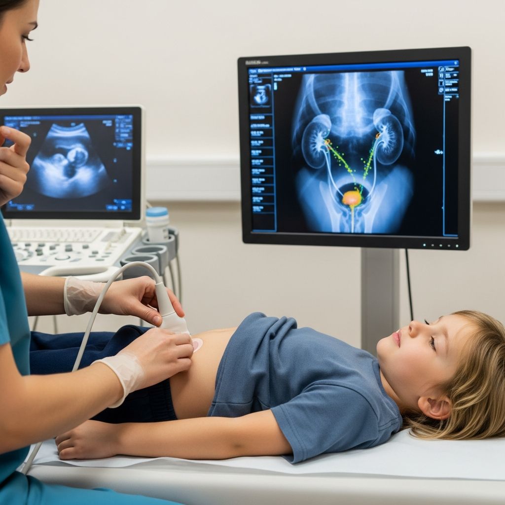
A voiding cystourethrogram, commonly abbreviated as VCUG, is a specialized X-ray test that helps doctors see how a child’s bladder and urethra work while the bladder is filling and when the child urinates. It is one of the main tests used to diagnose vesicoureteral reflux (urine flowing backward toward the kidneys) and other problems in the lower urinary tract.
This guide explains in clear language what the test is, when it is recommended, what your child will experience, and how to interpret the results, so you can approach the appointment with confidence.
What Is a VCUG and What Does It Show?
A VCUG is an imaging study that uses:
- X-rays or real-time X-ray video (fluoroscopy) to create pictures of the urinary tract.
- Contrast dye (a liquid that shows up clearly on X-ray images) placed into the bladder through a thin catheter.
During the test, the radiology team takes pictures while the bladder fills with the contrast liquid and again as the child urinates. This allows the doctor to see:
- The shape, size, and position of the bladder.
- How the urethra (tube that carries urine out) looks and functions.
- Whether urine and contrast are flowing backward from the bladder up toward the ureters and kidneys (vesicoureteral reflux).
- Any signs of blockage, narrowing, or structural abnormality in the urethra or lower urinary tract.
| Part of urinary tract | What the VCUG helps evaluate |
|---|---|
| Bladder | Size, shape, capacity, ability to hold and release urine, and any abnormal outpouchings or irregular walls. |
| Urethra | Whether the tube is open, narrowed, blocked, or has unusual curves or strictures that hinder urine flow. |
| Ureters/Kidneys | Whether urine or contrast flows backward toward the kidneys (reflux) when the bladder contracts. |
Why Might a Child Need a VCUG?
Doctors do not order a VCUG for every urinary problem. It is usually recommended when there is concern about how the urinary tract is built or how it functions. Common reasons include:
- Repeated urinary tract infections (UTIs), especially in infants and young children, to look for reflux or structural problems that might be causing infections.
- Known or suspected vesicoureteral reflux (VUR) seen on a previous test, to grade its severity and monitor changes over time.
- Abnormal prenatal ultrasound findings, such as swelling of the kidneys (hydronephrosis) or urinary tract dilation, that need more detailed evaluation after birth.
- Unusual urination patterns, such as very weak stream, difficulty starting urine, or dribbling, which may suggest a blockage or narrowing in the urethra.
- Follow-up after surgery on the urinary tract, to see whether the repair is working as intended and to check for residual reflux.
By identifying conditions like vesicoureteral reflux early, doctors can recommend the right treatment and help protect the child’s kidneys from long-term damage caused by repeated infections or high-pressure urine flow.
How the Urinary Tract Works (and Where VCUG Fits In)
To understand why this test is helpful, it is useful to know the main parts of the urinary tract:
- Kidneys – Filter waste and extra fluid from the blood to make urine.
- Ureters – Tubes that carry urine from each kidney down to the bladder.
- Bladder – A muscular storage sac that holds urine until it is time to urinate.
- Urethra – The tube that leads from the bladder to the outside of the body.
Normally, urine travels only one way: kidneys → ureters → bladder → urethra → outside. Valves at the points where the ureters meet the bladder help prevent urine from going back up toward the kidneys. In vesicoureteral reflux, these valves do not work properly, so urine can reverse direction when the bladder squeezes.
A VCUG lets doctors watch this process in real time. They can see how high the contrast travels, which side is affected, and how strongly it flows backward. This information helps classify reflux into grades from mild to severe and guides decisions about observation, medication, or surgery.
Step-by-Step: What Happens During a VCUG?
Although every hospital has its own routine, most VCUG tests follow a similar sequence. The exam is usually done in an outpatient radiology or imaging department, and most children go home right afterward.
1. Arrival and Check-In
- You and your child check in at the imaging department and complete consent and medical history forms.
- A technologist or nurse reviews your child’s medicines, allergies (especially to iodine or contrast dye), and recent illnesses.
- Pregnancy screening is done for teens who have begun menstruating because the test uses X-rays.
2. Getting Ready in the Imaging Room
- Your child usually changes into a hospital gown and removes metal objects like belts or jewelry that may interfere with images.
- Parents are often allowed to stay in the room wearing a protective lead apron, unless there is a specific reason they cannot.
- The staff explains the steps in child-friendly language, sometimes using dolls, pictures, or toys to demonstrate.
3. Placing the Catheter
The most uncomfortable part of the exam is the placement of a thin, flexible catheter into the urethra:
- The genital area is cleaned with an antiseptic solution to reduce the risk of infection.
- A small amount of lubricant is applied to the catheter to help it slide in more easily.
- The catheter is gently passed through the urethra into the bladder. Babies and young children are awake for this, and the process can sting or feel like pressure, but it is very brief.
- The catheter may be taped to the skin to help keep it in place during the exam.
In most cases, sedation is not required. Some centers may consider mild sedation for very anxious children or those with special needs, but this is decided individually and discussed in advance.
4. Filling the Bladder with Contrast
- Once the catheter is in place, contrast dye mixed with sterile liquid flows through the catheter into the bladder by gravity or a pump.
- Fluoroscopy (a live X-ray) is used to watch the bladder as it fills and to capture images at different stages.
- Your child may feel a growing sense of fullness or pressure as the bladder fills and may say they need to urinate.
5. Voiding (Urinating) During the X-ray
- When the bladder is full, the radiologist or technologist asks your child to urinate while lying on the table or sitting on a specialized container.
- Pictures are taken during urination to see whether contrast goes back up the ureters or if the urethra shows any narrowing or blockage.
- For babies in diapers, the team may encourage urination by gently stimulating or waiting for the natural urge, capturing images as the baby voids.
6. After the Images Are Complete
- Once enough images are obtained, the catheter is removed.
- A final set of pictures may be taken to confirm that the bladder has emptied properly.
- The whole procedure usually takes 30–45 minutes, depending on how quickly the bladder fills and the child can urinate.
Preparing Your Child Emotionally and Practically
Planning ahead can make the experience smoother and less frightening for both you and your child.
Before the Appointment
- Explain in simple terms what will happen: that doctors are taking special pictures of how pee comes out.
- Be honest that a small tube will be placed and may feel uncomfortable, but emphasize that you will be nearby.
- Ask if your child should come with a full or partially full bladder; specific instructions vary by center.
- Check whether any medicines should be paused before the test and report any allergies, especially to contrast material or iodine.
- Bring comfort items such as a favorite toy, blanket, tablet, or music to distract and soothe your child.
During the Test
- Stay calm and reassuring; children often mirror their parent’s emotional state.
- Hold your child’s hand, talk quietly, or sing to provide distraction.
- Follow the staff’s safety instructions, such as standing behind a shield or wearing a lead apron.
After the Test
- Your child can usually return to normal eating, drinking, and activities right away.
- Encourage extra fluids for the rest of the day to help flush out any remaining contrast and to dilute the urine.
- Urination may sting slightly the first few times afterward. A warm bath (if allowed by your provider) and plenty of fluids can help ease discomfort.
Risks, Safety, and Side Effects
VCUG is considered a low-risk procedure, but like any medical test, it has potential downsides. Understanding them can help you weigh benefits and risks with your child’s doctor.
- Radiation exposure: The test uses a small amount of ionizing radiation. Radiology teams follow pediatric safety principles to use the lowest dose needed for clear images.
- Discomfort or pain: Insertion of the catheter and bladder filling can cause brief stinging, pressure, or cramping, which stops soon after the test.
- Small risk of urinary tract infection: Because a catheter enters the bladder, there is a small chance of introducing bacteria. Many centers use sterile technique, and some children at higher risk may receive preventive antibiotics.
- Allergic reaction to contrast: Reactions to modern contrast agents used in the bladder are rare, but you should report any history of contrast or iodine allergy to the staff.
Call your child’s healthcare provider after the test if you notice:
- Fever or chills
- Burning or pain with urination that worsens or does not improve
- Difficulty urinating or inability to pass urine
- Blood in the urine that persists beyond a day
Understanding VCUG Results
A radiologist (a doctor specialized in imaging) reviews the pictures and sends a report to your child’s primary doctor or specialist. This process can take up to a couple of days, depending on the hospital’s workflow.
Results often address:
- Whether the bladder and urethra look normal in shape and size.
- Whether urine flows smoothly out of the bladder.
- Whether there is vesicoureteral reflux and, if so, how severe it is.
- Any evidence of blockage, narrowing, or outpouchings that may require further evaluation.
Reflux, if present, is usually graded from I (mild, affecting only the ureter) to V (severe, with significant kidney and ureter involvement). Treatment decisions depend on:
- The reflux grade
- Your child’s age
- How often UTIs happen
- Kidney function and any existing scarring
Possible management options include:
- Observation and repeat imaging to see if reflux improves with growth.
- Preventive antibiotics in some children to reduce the risk of infections.
- Behavioral and bladder training strategies for children who have voiding dysfunction.
- Surgical or minimally invasive procedures for children with severe or persistent reflux, or those who continue to have infections despite other treatments.
How VCUG Compares With Other Urinary Tract Tests
Your child may have additional imaging tests. Each serves a slightly different purpose.
| Test | What it shows | Key differences from VCUG |
|---|---|---|
| Renal and bladder ultrasound | Kidney size, shape, swelling, and basic bladder anatomy. | Uses sound waves (no radiation); does not show reflux during urination as clearly as VCUG. |
| Radionuclide cystogram | Presence and degree of reflux using a small amount of radioactive tracer. | Radiation dose may be lower but anatomic detail is less precise than VCUG; more often used for follow-up. |
| MAG3 or DMSA renal scan | Kidney function and scars or damage from past infections. | Focuses on kidney performance rather than direct visualization of the urethra and bladder while voiding. |
Frequently Asked Questions (FAQs)
At what age can children have a VCUG?
VCUG can be done in infants, children, and teenagers when medically needed, and even in adults if similar questions arise. Many VCUGs are performed in babies and toddlers after urinary tract infections or abnormal prenatal ultrasounds.
Will my child feel pain during the test?
The test is often described as uncomfortable but brief. The most bothersome moments are usually the catheter insertion and the feeling of a very full bladder. These sensations end quickly after the catheter is removed and the child urinates.
Is sedation or anesthesia necessary?
In most cases, no. VCUG is done with the child awake so that they can urinate on command. Some centers may use mild sedation for highly anxious children or those with special needs, but this is not routine and is considered carefully due to its own risks.
How soon will we get the results?
A radiologist reviews the images and sends a report to your child’s doctor, often within one to two business days. The timing depends on the hospital’s processes, so ask when to expect a follow-up call or visit.
Can my child go to school or daycare after a VCUG?
Most children can resume normal activities the same day, including school and play, as long as they feel well. Some may prefer to rest at home if they are tired or a bit uncomfortable with urination that day.
Does a normal VCUG mean my child will never get UTIs?
A normal VCUG rules out certain causes of UTIs, such as reflux or major structural abnormalities, but children can still develop infections for other reasons. Your child’s doctor may discuss other strategies, like hydration, timed voiding, and constipation management, to help reduce UTI risk.
References
- Voiding Cystourethrogram (VCUG): Procedure & Results — Cleveland Clinic. 2023-06-01. https://my.clevelandclinic.org/health/diagnostics/16431-vcug
- Pediatric VCUG (Voiding Cystourethrogram) — RadiologyInfo.org (ACR/RSNA). 2022-11-01. https://www.radiologyinfo.org/en/info/voidcysto
- Voiding cystourethrogram — MedlinePlus Medical Encyclopedia, U.S. National Library of Medicine. 2023-05-23. https://medlineplus.gov/ency/article/003784.htm
- Voiding Cystourethrogram (VCUG) — Children’s Hospital of Philadelphia. 2022-08-15. https://www.chop.edu/treatments/voiding-cystourethrogram-vcug
- Pediatric VCUG Test — Children’s Hospital Colorado. 2021-10-10. https://www.childrenscolorado.org/doctors-and-departments/departments/imaging-services/vcug-test/
- Voiding Cystourethrogram (VCUG) — Boston Children’s Hospital. 2021-05-18. https://www.childrenshospital.org/treatments/voiding-cystourethrogram