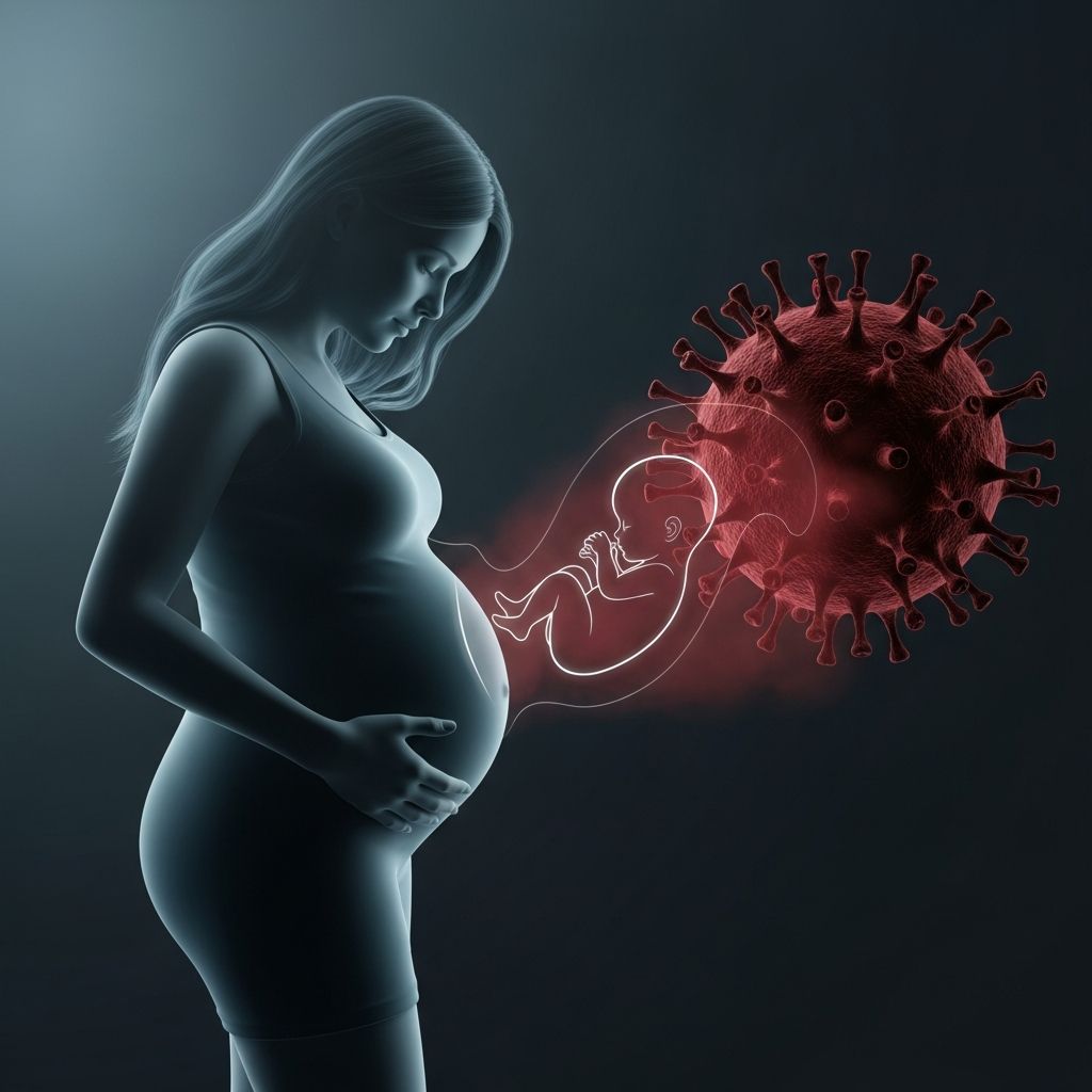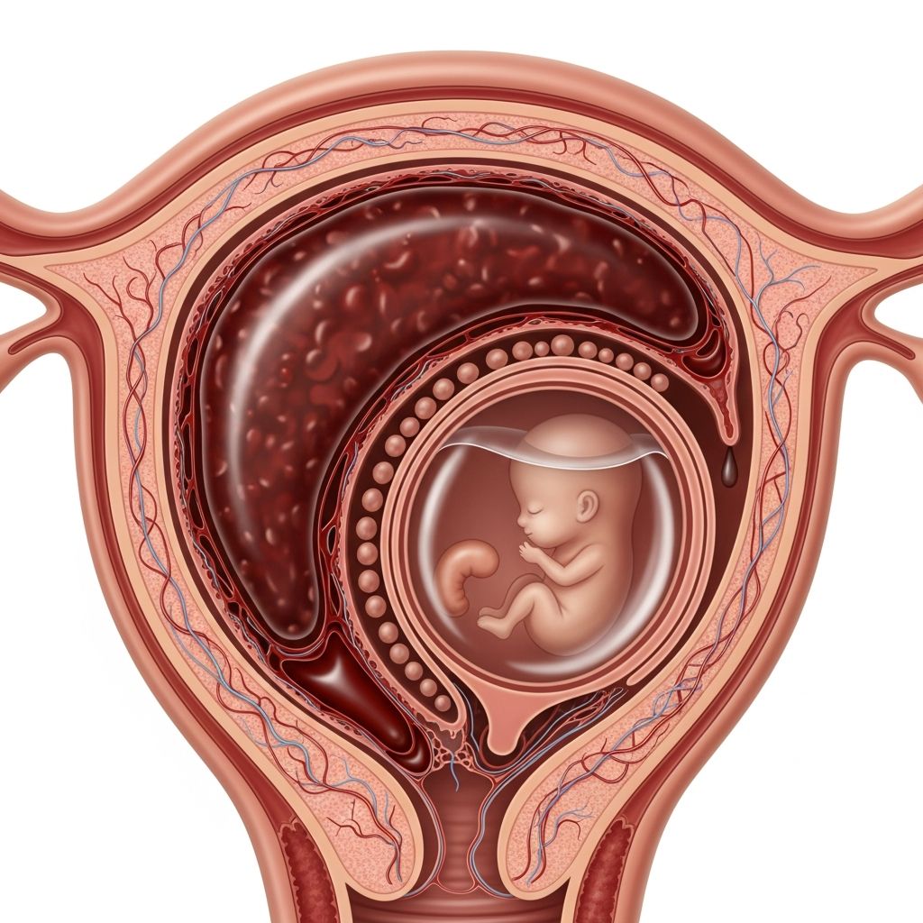
A subchorionic hematoma (SCH), also known as subchorionic hemorrhage or bleed, occurs when blood collects between the chorionic membrane and the uterine wall during pregnancy. This condition is frequently identified in the first trimester and represents a common cause of vaginal bleeding in early pregnancy.
Understanding the Anatomy and Formation of Subchorionic Hematoma
The chorion is the outermost membrane surrounding the amniotic sac, anchoring the developing placenta to the uterus. When small blood vessels in this area rupture due to placental separation or fragility, blood accumulates in the space between the chorion and the decidua (the uterine lining), forming a hematoma. This collection appears as a crescent-shaped, hypoechoic area on ultrasound imaging, varying in size from small spots to larger masses.
Pathogenesis often stems from impaired trophoblast invasion during early placental development, leading to fragile vessels and bleeding along the path of least resistance. Factors like maternal hypercoagulability or immune imbalances, such as Th1/Th2 cytokine shifts, may contribute to vessel contraction and clot formation in the subchorionic space.
Recognizing the Signs and Symptoms
The hallmark symptom of SCH is vaginal bleeding, which can range from light spotting to heavy flow with clots, typically in the first half of pregnancy. Some individuals experience pelvic cramping or lower abdominal discomfort, though many cases are asymptomatic and discovered incidentally during routine ultrasounds.
- Light spotting: Often pink or brown, resembling implantation bleeding.
- Heavy bleeding: May include clots, mimicking miscarriage symptoms.
- Associated pains: Mild belly ache, back pain, or tenderness that persists.
- No symptoms: Common in smaller hematomas detected only via imaging.
Differentiating SCH from other bleeding causes is crucial, as symptoms overlap with implantation, infections, cervical changes, ectopic pregnancy, or miscarriage.
Diagnostic Approaches and Ultrasound Insights
Diagnosis relies primarily on transvaginal ultrasound, the gold standard for visualizing SCH. It appears as a dark fluid collection adjacent to the gestational sac, often crescent or triangular in shape. Key ultrasound features include irregular texture or speckled amniotic fluid, distinguishing it from normal membrane non-fusion before 16 weeks.
Healthcare providers assess hematoma size relative to the gestational sac: small (<20% of sac circumference), medium (20-50%), or large (>50%), as size correlates with potential risks. Follow-up scans monitor resolution, as most SCH shrink and resolve spontaneously.
| Hematoma Size | Description | Typical Resolution Time |
|---|---|---|
| Small | <20% of sac | 2-8 weeks |
| Medium | 20-50% of sac | 4-12 weeks |
| Large | >50% of sac | Variable, higher monitoring |
Serial ultrasounds every 1-2 weeks help track progression, especially if bleeding persists.
Potential Causes and Risk Factors
The exact etiology remains unclear, but several factors increase susceptibility:
- Uterine abnormalities: Malformations affecting implantation.
- Prior pregnancy losses: History of recurrent miscarriages.
- Infections: Pelvic or cervical infections weakening tissues.
- Trauma: Physical injury to the abdomen.
- Hypertensive disorders: Early preeclampsia or severe hypertension.
- Assisted reproduction: IVF pregnancies show higher incidence.
- Coagulation issues: Maternal hypercoagulable states leading to thrombosis.
Rare triggers include drug abuse (e.g., cocaine) or thrombolytic therapy.
Associated Risks and Complications
While most SCH resolve without issue, larger hematomas or those persisting beyond the first trimester elevate risks.
- Miscarriage: Increased odds, especially with large SCH (up to 5-10% higher loss rate before 24 weeks).
- Preterm birth: 18-20% risk in ongoing pregnancies.
- Preeclampsia: Potential link due to vascular fragility.
- Fetal growth restriction: Reduced nutrient supply from placental issues.
- Placental abruption: Rare but serious separation later in pregnancy.
Research is mixed; some studies find no elevated adverse outcomes, while others confirm associations, particularly with hematoma size and location.
Management and Treatment Strategies
Treatment is primarily conservative, focusing on rest and monitoring, as most SCH resolve naturally.
- Bed rest: Pelvic rest advised, avoiding intercourse, heavy lifting, or strenuous activity.
- Progesterone supplementation: May support uterine lining in threatened abortion cases.
- Follow-up ultrasounds: To confirm resolution.
- Pain management: Acetaminophen for cramps; avoid NSAIDs.
- Hospitalization: Rare, for heavy bleeding or hemodynamic instability.
No surgical interventions are standard; aspiration risks further bleeding.
Lifestyle Adjustments for Expectant Mothers
To minimize complications:
- Stay hydrated and eat a balanced diet rich in iron and folate.
- Avoid smoking, alcohol, and recreational drugs.
- Monitor fetal movements once viable.
- Report worsening symptoms immediately: heavy bleeding, severe pain, fever, or dizziness.
Emotional support is vital; anxiety from bleeding is common—consider counseling.
When to Seek Immediate Medical Attention
Any pregnancy bleeding warrants prompt evaluation to rule out ectopic pregnancy, molar pregnancy, or abruption. Emergency signs include:
- Heavy bleeding soaking a pad hourly.
- Severe abdominal pain or shoulder tip pain.
- Dizziness, fainting, or rapid heartbeat.
- Fever or foul-smelling discharge indicating infection.
Contact your obstetrician or visit the ER without delay.
Prognosis and Long-Term Outlook
Over 90% of small SCH resolve by the second trimester with healthy outcomes. Even larger ones often lead to term deliveries with vigilant care. Post-resolution, routine prenatal care resumes, with no lasting effects on future pregnancies unless recurrent.
Frequently Asked Questions About Subchorionic Hematoma
Can subchorionic hematoma cause miscarriage?
Yes, larger hematomas increase miscarriage risk, particularly in the first trimester, but many pregnancies continue successfully with monitoring.
Is bed rest necessary for SCH?
Pelvic rest is commonly recommended to reduce bleeding, though strict bed rest evidence is limited—follow provider guidance.
How long does it take for SCH to resolve?
Small ones often disappear in 2-8 weeks; larger may take longer, monitored via ultrasound.
Does SCH affect the baby?
Rarely, if it resolves early; risks like growth restriction arise only with complications.
Can I have sex with SCH?
No, pelvic rest prohibits intercourse until resolution to avoid aggravating the hematoma.
References
- Subchorionic bleeding: Causes, symptoms, and risks — Medical News Today. 2023-10-12. https://www.medicalnewstoday.com/articles/323307
- Subchorionic Hematoma: Causes, Symptoms & Treatment — Cleveland Clinic. 2023-08-15. https://my.clevelandclinic.org/health/symptoms/23511-subchorionic-hematoma
- Subchorionic hematoma: Research status and pathogenesis (Review) — PMC / NIH. 2024-02-20. https://pmc.ncbi.nlm.nih.gov/articles/PMC10865452/
- Subchorionic Hematoma — Sharp Mary Birch Maternal Fetal Medicine. 2022-11-05. https://sandiegoperinatal.com/patient-education/pregnancy-complications/subchorionic-hematoma/
- What You Should Know About Subchorionic Hematoma in Pregnancy — WebMD. 2024-01-10. https://www.webmd.com/baby/what-is-a-subchorionic-hematoma