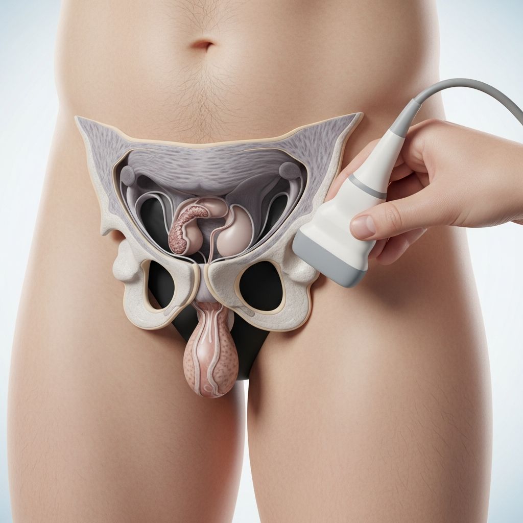
A scrotal ultrasound is a safe, noninvasive imaging test that uses high-frequency sound waves to create detailed pictures of a boy’s testicles, epididymis, and surrounding scrotal tissues. Parents often seek information when their child experiences scrotal pain, swelling, or a noticeable lump, as this procedure helps doctors accurately diagnose potential issues without radiation exposure.
Understanding the Scrotum and Its Importance in Boys
The scrotum is the sac of skin that holds the testicles, which are crucial for producing sperm and hormones like testosterone. Nearby structures include the epididymis—coiled tubes where sperm mature—and blood vessels. In children and teens, these areas can develop problems due to injury, infection, or developmental issues. Early detection through imaging is vital to prevent complications like infertility or tissue damage.
Boys may notice symptoms such as sudden sharp pain, gradual swelling, or a firm bump. These warrant a doctor’s visit, where physical exams might lead to recommending a scrotal ultrasound for clearer insights.
Key Reasons Doctors Recommend Scrotal Ultrasound in Children
This imaging modality serves multiple diagnostic purposes tailored to pediatric needs. Here’s why it’s commonly ordered:
- Assessing lumps or masses: Determines if a scrotal lump is a fluid-filled cyst or a solid tumor, guiding treatment decisions.
- Evaluating pain or swelling: Identifies inflammation (epididymitis or orchitis), abscesses, or other causes of discomfort.
- Detecting testicular torsion: A medical emergency where the testicle twists, cutting off blood flow; ultrasound confirms blood flow absence.
- Checking for varicocele: Enlarged veins that may affect fertility later; common in teens.
- Post-trauma evaluation: Reveals hematomas, ruptures, or other injuries from sports or accidents.
- Locating undescended testes: Helps find cryptorchid testicles in the abdomen or groin.
In pediatric cases, distinguishing between surgical emergencies like torsion and benign conditions is critical, as delays can lead to testicle loss.
How Scrotal Ultrasound Technology Works
Unlike X-rays, ultrasound employs sound wave echoes, similar to sonar used by submarines. A handheld probe (transducer) emits high-frequency waves that travel through gel applied to the skin, bounce off tissues, and return as echoes. A computer interprets these to form real-time images on a screen, showing structures, movement, and blood flow via Doppler mode.
For scrotal scans, a high-frequency linear transducer (7.5-10 MHz) provides sharp resolution of small parts like testicles. This allows visualization of tissue texture—solid vs. cystic—and vascularity, essential for accurate diagnosis.
Step-by-Step: What Happens During Your Child’s Scrotal Ultrasound
The procedure is quick, typically 15-30 minutes, and performed in a radiology suite. No sedation is usually needed for children who can stay still.
- Preparation: Your child changes into a gown. The technologist explains the process to reduce anxiety.
- Positioning: Lying on their back with legs spread, a towel or tape elevates the scrotum for better imaging. The unaffected side is scanned first for comparison.
- Gel application: Warm, water-based gel is spread on the scrotum to conduct sound waves without air interference.
- Scanning: The sonographer glides the probe in longitudinal and transverse planes, pressing gently. Your child may feel mild pressure but no pain. Images and videos are captured.
- Doppler assessment: Color flow checks blood supply symmetry.
- Completion: Gel is wiped off; your child dresses and resumes activities immediately.
Parents can stay present for comfort, encouraging stillness.
Preparation Tips for Parents and Children
No special prep like fasting is required. Advise loose clothing for easy access. Discuss any allergies or sensitivities beforehand. For younger boys, use simple explanations: “It’s like a camera using jelly and a wand to take pictures inside—no needles!” Practice deep breathing to stay calm.
| Age Group | Tips |
|---|---|
| Under 5 | Use storybooks about doctor visits; bring a favorite toy. |
| 5-10 | Show ultrasound videos; emphasize it’s quick and painless. |
| Teens | Explain medical reasons honestly; respect privacy with chaperone options. |
Safety Profile: Benefits Outweigh Minimal Risks
Scrotal ultrasound is exceptionally safe—no ionizing radiation, needles, or anesthesia. Mild discomfort from probe pressure is rare and temporary. Benefits include precise diagnosis enabling timely interventions, cost-effectiveness, and real-time imaging for procedures like biopsies.
Risks are negligible: no known long-term effects from sound waves at diagnostic levels. It’s preferred over CT or MRI for initial scrotal evaluations.
Interpreting Results: What Parents Should Know
A radiologist analyzes images, sending a report to your doctor within hours or days. Normal findings show uniform testicles with good blood flow. Abnormalities might include:
- Hydrocele: Fluid around testicle (often benign).
- Varicocele: Vein dilation, monitored or surgically corrected.
- Torsion: Absent flow requiring surgery.
- Masses: Further tests like biopsy if solid.
Follow-up depends on findings—watchful waiting for minor issues or urology referral for others.
When to Seek Urgent Scrotal Ultrasound
Rush to ER if your child has sudden severe pain, nausea, vomiting, or scrotal redness/swelling—these signal torsion or strangulated hernia. Routine cases follow outpatient scheduling.
Common Parental Concerns Addressed
Is it painful? Generally no; brief pressure at most.
Does it affect fertility? No evidence; it’s diagnostic only.
What if my child is embarrassed? Technologists are trained for sensitivity; parents accompany.
Frequently Asked Questions (FAQs)
Why might my young son need a scrotal ultrasound?
It evaluates pain, swelling, lumps, or trauma to prevent serious issues like torsion.
How long do results take?
Preliminary same-day; full report in 1-2 days.
Can girls or toddlers have similar issues?
This is male-specific; labial concerns use different imaging.
Is sedation ever used?
Rarely for uncooperative young children; discuss alternatives.
Follow-up after normal scan?
Usually none, but monitor symptoms.
This guide empowers parents with knowledge for informed decisions on scrotal health. Consult your pediatrician for personalized advice.
References
- Ultrasound Imaging of the Scrotum — RadiologyInfo.org. 2023. https://www.radiologyinfo.org/en/info/us-scrotal
- Scrotal ultrasound — FloridaHealthFinder. 2024. https://quality.healthfinder.fl.gov/health-encyclopedia/HIE/1/003780
- Scrotal Ultrasonography – Medical Clinical Policy Bulletins — Aetna. 2025-01-15. https://www.aetna.com/cpb/medical/data/500_599/0532.html
- Testicular Ultrasound: Purpose, Risks, Procedure, and More — Healthline. 2023-05-10. https://www.healthline.com/health/testicle-ultrasound
- Small Parts – Testicular Ultrasound — American College of Emergency Physicians (ACEP). 2024. https://www.acep.org/sonoguide/advanced/testicular
- Testicular Scan: What Should You Expect? — Harley Street Ultrasound. 2024. https://www.harleystreetultrasound.com/articles/what-is-a-testicular-ultrasound/
- Testicular Ultrasound: Purpose, Procedure, Risks & Results — Cleveland Clinic. 2024-02-20. https://my.clevelandclinic.org/health/diagnostics/testicular-ultrasound