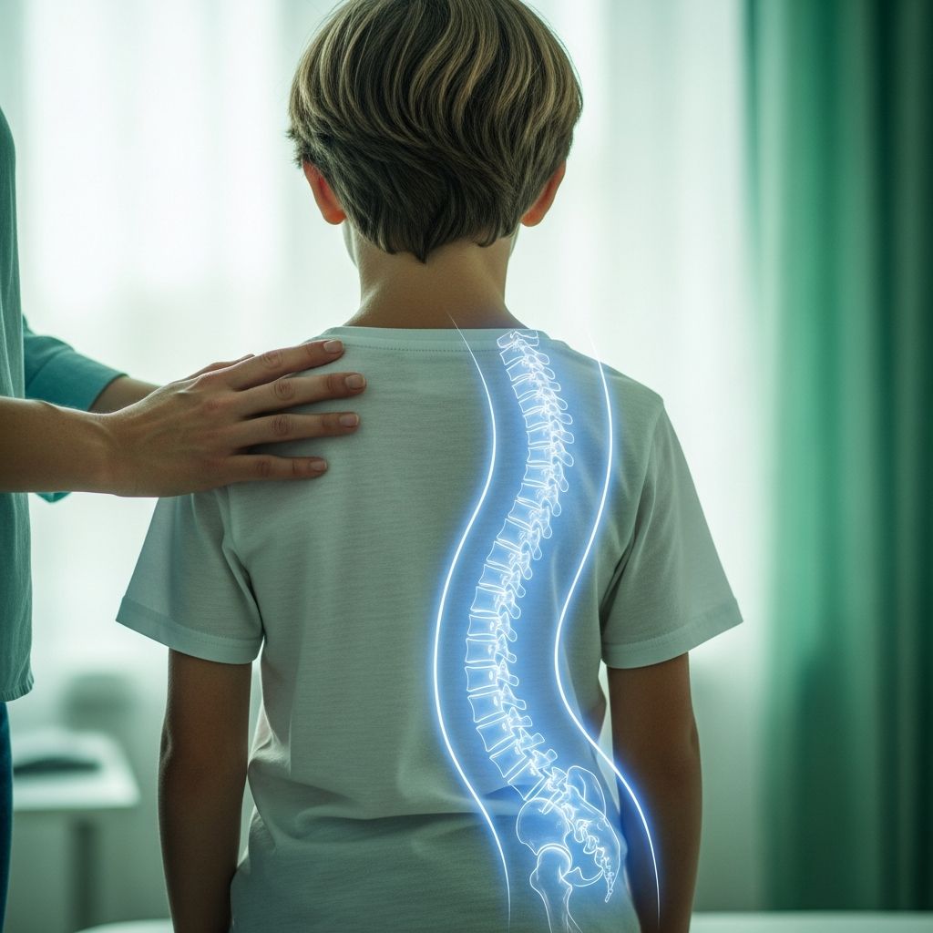
Scoliosis X-rays are the cornerstone of diagnosing and managing spinal curvature in children, allowing doctors to measure the extent of the curve and track its progression accurately.
Understanding Scoliosis Basics
Scoliosis involves an abnormal sideways curvature of the spine, often detected during childhood growth spurts. A curve exceeding 10 degrees on an X-ray confirms the diagnosis, distinguishing it from normal spinal variations. This condition affects millions of children worldwide, with most cases being idiopathic, meaning no clear cause is identified, though growth phases can exacerbate it.
Early detection through school screenings or routine check-ups is vital, as curves can worsen rapidly during puberty. Parents may notice uneven shoulders, hips, or a prominent rib hump when their child bends forward. These visual cues prompt further imaging to quantify the issue.
The Essential Role of X-Rays in Diagnosis
X-rays provide clear images of the spine’s alignment, capturing views from the back (posteroanterior or PA) and side (lateral) while the child stands upright for the most precise assessment. This positioning reveals the true degree of curvature, rotation, and any associated vertebral irregularities.
Unlike physical exams, which spot asymmetries, X-rays offer measurable data. They help classify scoliosis types, such as adolescent idiopathic (common in teens), congenital (present at birth), or neuromuscular (linked to conditions like cerebral palsy).
- Standing posture simulates daily spinal load for realistic curve evaluation.
- Full-spine views cover thoracic (mid-back), lumbar (lower back), and sometimes cervical regions.
- Lead shields protect sensitive organs like the thyroid, breasts, and gonads during exposure.
Key Measurements from Scoliosis X-Rays
Radiologists analyze X-rays using standardized tools to guide treatment. The Cobb angle, the gold standard, measures the curve’s severity by drawing lines along the tilted vertebrae and calculating the intersection angle.
| Cobb Angle Range | Severity Level | Typical Management |
|---|---|---|
| 10-24 degrees | Mild | Observation, possible bracing |
| 25-39 degrees | Moderate | Bracing, physical therapy |
| 40+ degrees | Severe | Surgery consideration |
Other features include the Risser sign, which assesses skeletal maturity via iliac crest ossification on pelvic X-rays, predicting progression risk. Rotation is evaluated through vertebral pedicle offset or rib hump prominence.
Bone age X-rays of the hand or wrist examine growth plate closure, crucial since scoliosis advances most during rapid growth.
Advanced Imaging: Minimizing Radiation Risks
Traditional X-rays use ionizing radiation, raising concerns for growing children who may need serial imaging every 4-6 months. Cumulative exposure from frequent scans can pose long-term risks, prompting innovations like EOS imaging.
EOS, a slot-scanning technology, delivers 6-9 times less radiation while providing 3D spine models from biplanar (front and side) images in standing position. This enhances accuracy for brace fitting and surgical planning without excess dose.
Many specialized centers prioritize EOS or other low-dose protocols. MRI complements X-rays for non-radiation soft tissue views, detecting tethered cords, tumors, or syrinxes underlying atypical scoliosis, especially in young children.
Preparing Your Child for a Scoliosis X-Ray
Preparation is straightforward: wear loose clothing, avoid metal jewelry, and arrive with empty bladder for comfort. Explain the quick process (2-5 minutes) to reduce anxiety—it’s painless, like standing still for a photo.
Technicians use gentle restraints if needed for young kids and provide gowns. Parental presence calms children; discuss any claustrophobia or prior fears beforehand. Results are typically reviewed same-day by a specialist.
- Schedule during less busy times to minimize wait.
- Bring a favorite toy or snack post-scan.
- Ask about EOS availability upfront.
Interpreting Your Child’s X-Ray Results
Reports detail Cobb angle, curve location (e.g., thoracic vs. lumbar), rotation, and maturity indicators. A 15-degree thoracic curve in a growing 12-year-old might warrant bracing, while a stable 20-degree curve post-puberty could just need monitoring.
Progression is tracked by comparing serial X-rays; a 5-degree increase signals intervention. Understand terms: ‘apex’ (most curved vertebra), ‘end vertebrae’ (curve boundaries), and stability factors like balanced shoulders.
Consult a pediatric orthopedist for personalized insights—family history and growth velocity influence decisions.
Treatment Pathways Informed by X-Rays
X-ray data drives options: observation for mild curves, bracing (e.g., Boston brace) for 20-40 degrees in growing kids to halt progression, or surgery (spinal fusion) for severe cases.
Post-treatment X-rays verify brace efficacy or surgical correction, often using low-dose EOS for follow-ups. Early imaging enables conservative approaches, improving long-term posture and lung function.
Frequently Asked Questions (FAQs)
Is a single X-ray enough to diagnose scoliosis?
No, initial X-rays confirm the curve, but follow-ups monitor changes, especially during growth spurts.
How often will my child need scoliosis X-rays?
Every 4-6 months if growing and at risk; less frequently if stable. Low-dose options reduce concerns.
Does X-ray radiation harm my child?
Risks are low with modern protocols; EOS cuts exposure significantly compared to traditional methods.
What if the X-ray shows no curve over 10 degrees?
It rules out scoliosis; asymmetries may stem from posture or muscle imbalance, warranting observation.
Can home exercises replace X-rays?
No, imaging is essential for precise measurement; exercises support but don’t diagnose.
Empowering Parents: Next Steps After Imaging
Armed with X-ray insights, partner with specialists. Seek second opinions at scoliosis centers, join support groups, and track growth charts. Regular monitoring catches changes early, optimizing outcomes like straight spines and active lifestyles.
Advocate for low-radiation tech and holistic care, including nutrition for bone health. With vigilant imaging, most children thrive without surgery.
References
- Scoliosis – Diagnosis and treatment — Mayo Clinic. 2023-10-01. https://www.mayoclinic.org/diseases-conditions/scoliosis/diagnosis-treatment/drc-20350721
- Navigating Scoliosis Xrays: A Parent’s Essential Guide — Treating Scoliosis. 2024-05-15. https://treatingscoliosis.com/blog/scoliosis-x-ray-a-guide-for-parents/
- Scoliosis in Children and Teenagers: An Overview — Hospital for Special Surgery (HSS). 2023-08-20. https://www.hss.edu/health-library/conditions-and-treatments/scoliosis-in-children-teenagers
- Diagnosing Scoliosis in Children — NYU Langone Health. 2024-02-10. https://nyulangone.org/conditions/scoliosis-in-children/diagnosis
- Scoliosis in Children and Teens: Diagnosis, Treatment, and Steps to Take — National Institute of Arthritis and Musculoskeletal and Skin Diseases (NIAMS), NIH. 2023-11-05. https://www.niams.nih.gov/health-topics/scoliosis/diagnosis-treatment-and-steps-to-take
- Scoliosis X-Ray: Key Insights for Patients & Families — ScoliCare. 2024-01-22. https://scolicare.com/denver-colorado/scoliosis-x-ray-key-insights-for-patients-families/
- EOS Imaging Reduces Radiation Exposure for Children with Scoliosis — Duke Health. 2023-06-12. https://www.dukehealth.org/blog/eos-imaging-reduces-radiation-exposure-children-scoliosis