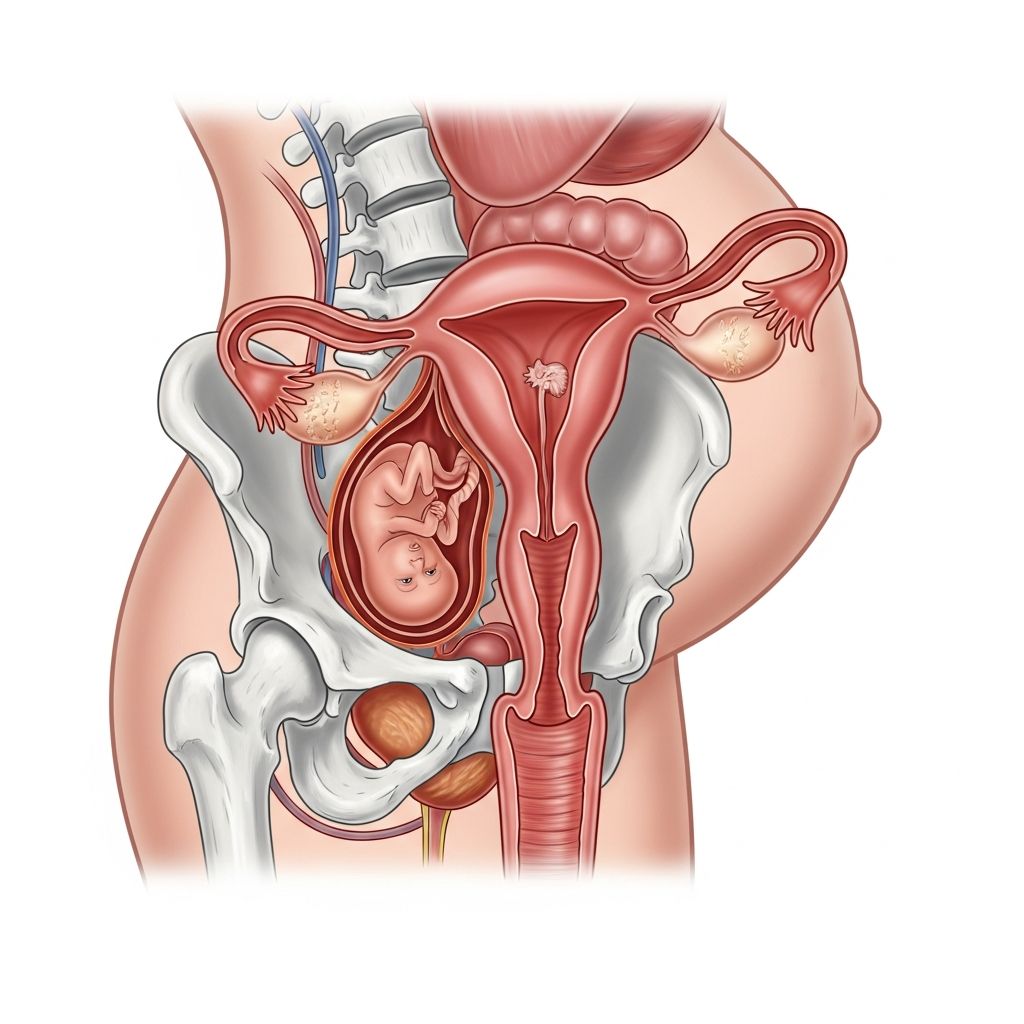
Understanding Uterine Retroversion: A Natural Anatomical Variation
Pregnancy brings countless physical changes, many of which expectant mothers anticipate with excitement and wonder. However, for some women, the appearance of their baby bump follows an unexpected timeline. One significant factor influencing this variation is the position of the uterus itself. A substantial portion of the female population naturally has what medical professionals call a retroverted or tilted uterus—a condition where this vital reproductive organ tilts backward toward the spine rather than maintaining the more common forward-facing orientation.
This anatomical difference is far more common than many women realize. Approximately 15 to 25 percent of women have a naturally tilted uterus, making it a variation rather than an abnormality. In many ways, having a retroverted uterus is comparable to being left-handed in a predominantly right-handed world—it represents a normal human variation in anatomy rather than a medical concern requiring intervention.
The Mechanics of Uterine Positioning and Baby Bump Development
To understand how uterine retroversion affects the visible baby bump, it helps to first comprehend normal uterine anatomy and pregnancy progression. In their standard configuration, most uteri point slightly forward, angling toward the bladder and anterior abdominal wall. This anterior positioning means that as pregnancy advances and the uterus expands, the growth naturally projects outward, creating the characteristic rounded belly that becomes visible relatively early in pregnancy.
When a uterus is tilted backward, it initially grows within the pelvis rather than extending into the abdominal cavity. During the first trimester and often into the second, the expanding uterus occupies pelvic space rather than creating a visible protrusion through the abdominal wall. This spatial difference explains why some pregnant women appear to “show” much later than their peers, sometimes not displaying a noticeable bump until four, five, or even six months into pregnancy.
The good news for expectant mothers with retroversion is that this pattern typically reverses naturally. As pregnancy progresses and the fetus grows larger, the expanding uterus gradually tilts forward and rises out of the pelvis into the abdominal cavity. By the second trimester, usually around 12 to 13 weeks, most women with retroverted uteri experience this spontaneous positional shift, after which their baby bumps become increasingly visible.
What Causes a Tilted Uterus?
Uterine retroversion can develop through several distinct pathways. Understanding these causes helps demystify why some women are born with this variation while others develop it later in life.
Developmental and Natural Causes
Some women are born with a naturally retroverted uterus as a result of their individual anatomical development. During fetal development and childhood, the uterus gradually shifts into its adult position. In some individuals, this repositioning never fully occurs, leaving the uterus in a tilted orientation. This represents a normal developmental variation with no pathological basis.
Post-Surgical Complications and Scarring
Pelvic surgical procedures frequently contribute to uterine retroversion. When surgical sites heal, they can produce adhesions—bands of scar tissue that abnormally join anatomical structures that should remain separate. These adhesions can attach the uterus to nearby organs or pelvic structures, physically pulling it into a backward tilt. Common surgeries that may result in adhesions include cesarean delivery, dilation and curettage procedures, and other gynecological interventions.
Endometriosis and Inflammatory Conditions
Endometriosis, a condition where tissue similar to the uterine lining grows outside the uterus, frequently causes retroversion. The abnormal tissue growth can create adhesions that bind the uterus to surrounding structures, pulling it backward. Additionally, pelvic inflammatory disease and other chronic infections can produce scarring that contributes to uterine tilting.
Uterine Fibroids and Structural Growth
Benign growths within or on the uterus, known as fibroids, can distort uterine shape and position. Large fibroids can shift the uterus’s center of gravity, causing it to tilt backward. These non-cancerous growths affect a significant percentage of women and represent another common cause of retroversion.
Post-Pregnancy Changes
Pregnancy and childbirth place substantial stress on the pelvic support structures. The ligaments holding the uterus in place stretch significantly during gestation and labor. While these ligaments typically regain their tone after delivery, sometimes they remain lax or weakened. This reduced support can allow the uterus to settle into a retroverted position postpartum.
Hormonal and Age-Related Factors
As women transition through menopause, declining estrogen levels weaken the muscles and ligaments supporting pelvic organs. This hormonal shift can cause previously forward-tilted uteri to gradually tilt backward. Age-related changes in pelvic tissue elasticity contribute to increased retroversion prevalence in older women.
Recognizing Symptoms and Clinical Presentation
While many women with retroverted uteri experience no symptoms whatsoever, others report specific complaints that may vary in intensity and frequency.
- Dyspareunia: Pain or discomfort during sexual intercourse represents one of the most commonly reported symptoms, particularly during deep penetration
- Menstrual complications: Some women experience intensified menstrual cramping and prolonged or heavier periods
- Urinary challenges: Difficulty with bladder emptying and recurrent urinary tract infections affect some individuals
- Tampon insertion difficulties: The tilted uterine position can make inserting tampons more challenging or uncomfortable
- Pelvic discomfort: Generalized pelvic pressure or aching, particularly during menstruation, occurs in some cases
It’s important to emphasize that many women with retroverted uteri experience none of these symptoms and discover their uterine position only incidentally during imaging for other reasons or during pregnancy.
Pregnancy Considerations and Fetal Development
For most women with retroverted uteri, pregnancy progresses without complications. The delayed visibility of the baby bump represents the primary noticeable difference compared to women with forward-tilted uteri. However, healthcare providers should be aware of several specific pregnancy-related considerations.
First Trimester Assessment Challenges
During initial prenatal evaluations, physicians may experience difficulty locating and visualizing the fetus on physical examination during the first three months. Ultrasound imaging typically resolves this challenge, but the backward uterine position can make manual palpation and auscultation more difficult than expected. This represents a temporary technical challenge rather than a medical problem.
The Natural Postural Transition
One of the most remarkable aspects of pregnancy with retroversion is the spontaneous positional shift that occurs as gestation advances. The expanding uterus, now containing an increasingly large fetus, naturally and gradually rotates forward and lifts out of the pelvis. This physiological accommodation allows adequate space for continued fetal growth and proper positioning for delivery. Most women experience this transition between weeks 12 and 14, though timing varies individually.
Rare Complications: Uterine Incarceration
In a small percentage of cases, the retroverted uterus does not spontaneously flip forward as expected during pregnancy. Instead, it becomes “snagged” or caught on pelvic bones, typically the sacrum. This uncommon condition, termed incarcerated uterus, usually manifests between weeks 12 and 14 with symptoms including pelvic pain and urinary retention. Prompt medical attention can resolve this condition, and it does not necessarily compromise pregnancy viability or fetal health.
Managing Retroversion: Treatment Options and Interventions
Treatment approaches for retroverted uterus vary based on the underlying cause, symptom severity, and individual circumstances. Many women require no treatment whatsoever, particularly if asymptomatic.
Conservative Management Strategies
For women experiencing mild symptoms, conservative approaches often prove effective. Pelvic floor exercises, including Kegel exercises, can strengthen the muscles supporting the uterus and may help reposition or stabilize it. Physical therapy specifically targeting pelvic anatomy can provide additional benefits. Heat application and anti-inflammatory medications address pain symptoms when needed.
Pessary Devices
A pessary represents a non-surgical intervention option. This small, plastic, donut-shaped device is inserted into the vagina where it provides physical support, effectively propping the uterus forward. Pessaries work well for some women but require regular cleaning and monitoring for potential complications. They can be removed at any time and do not permanently alter anatomy.
Treating Underlying Conditions
When retroversion results from endometriosis, fibroids, or adhesions, addressing these underlying conditions often improves uterine positioning. Surgical treatment of endometriosis, fibroid removal, and adhesion lysis can sometimes allow the uterus to resume a more anterior position. Managing these conditions provides benefits beyond uterine positioning alone.
Surgical Correction
Surgical procedures to reposition the uterus remain available though are rarely recommended. These procedures carry surgical risks including infection, bleeding, and formation of new adhesions. Most healthcare providers reserve surgical correction for cases where symptoms significantly impact quality of life and conservative measures have failed.
Fertility and Reproductive Implications
A frequently asked question concerns whether retroversion affects fertility or the ability to conceive. The straightforward answer is that retroversion alone does not cause infertility. Women with retroverted uteri conceive and carry pregnancies successfully at rates comparable to the general population.
However, if retroversion results from underlying conditions such as endometriosis or pelvic inflammatory disease, these conditions themselves may impact fertility. In such cases, treating the underlying disorder rather than the retroversion itself improves reproductive outcomes. The distinction between retroversion as a simple anatomical variant versus retroversion secondary to pathology becomes clinically important for fertility planning.
Frequently Asked Questions
Will my baby bump eventually become visible if I have a retroverted uterus?
Yes, in the vast majority of cases, your baby bump will become increasingly visible as pregnancy progresses. Most women with retroversion experience their uterus naturally flipping forward between weeks 12 and 14, after which the bump becomes apparent much like in women with forward-tilted uteri.
Does retroversion increase my risk of miscarriage?
No, retroversion does not increase miscarriage risk. Pregnancy loss occurs at similar rates regardless of uterine position. Retroversion is a positional variation, not a pathological condition affecting pregnancy viability.
Can retroversion affect how I deliver my baby?
Uncomplicated retroversion does not affect delivery method or outcomes. Your healthcare provider may plan labor and delivery based on overall pregnancy progression and other individual factors, but retroversion alone does not necessitate cesarean delivery or other modifications.
Should I tell my healthcare provider about my retroversion?
Yes, if you know you have a retroverted uterus, informing your prenatal care team helps them anticipate potential assessment challenges in early pregnancy and monitor for rare complications like incarcerated uterus. This knowledge allows for more individualized and attentive care.
Is retroversion permanent or can it resolve on its own?
Retroversion can be permanent, particularly if it represents a natural anatomical variation. However, if it resulted from causes like adhesions or inflammation, treating those underlying conditions may allow the uterus to resume a forward position. Pregnancy itself temporarily changes uterine position for most women.
Moving Forward with Confidence
Understanding that a retroverted uterus represents a normal anatomical variation rather than a medical problem forms an important foundation for approaching pregnancy and reproductive health with confidence. While the delayed visibility of a baby bump during early pregnancy can generate concerns or unwanted commentary from those unfamiliar with retroversion, knowing that this pattern is expected and temporary helps contextualize the experience. Most women with retroverted uteri have uncomplicated pregnancies, deliver healthy babies, and experience no long-term reproductive consequences from their anatomical variation. Open communication with healthcare providers, combined with accurate information about what to expect, ensures that pregnancy progresses smoothly despite this common anatomical difference.
References
- What Is a Retroverted Uterus? — Cleveland Clinic. 2024. https://my.clevelandclinic.org/health/diseases/23426-retroverted-uterus
- Tipped Uterus: Tilted Uterus — American Pregnancy Association. https://americanpregnancy.org/getting-pregnant/tipped-uterus/
- Retroverted Uterus — Better Health Channel, Department of Health, State Government of Victoria, Australia. 2024. https://www.betterhealth.vic.gov.au/health/conditionsandtreatments/retroverted-uterus
- What Is a Tilted or Retroverted Uterus? — The Bump. https://www.thebump.com/a/tilted-uterus-during-pregnancy
- A Woman’s Baby Bump Grew Backwards Due to a Retroverted Uterus — Business Insider. 2017. https://www.businessinsider.com/womans-baby-bump-grows-backwards-retroverted-uterus-2017-12
- Retroverted Uterus & Fertility: Causes, Symptoms & Treatment Options — CCRM Fertility. 2024. https://www.ccrmivf.com/blog/retroverted-uterus-fertility-causes-symptoms-treatment/