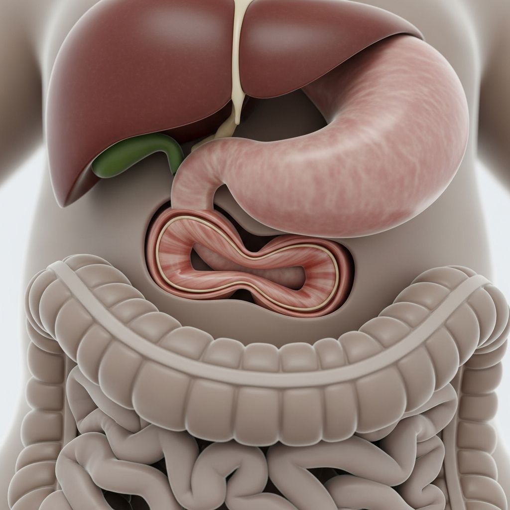
Understanding Pyloric Stenosis in Newborns and Young Infants
Pyloric stenosis represents a significant medical condition affecting infants during their earliest months of life. This disorder occurs when the muscle controlling the passage between the stomach and small intestine becomes abnormally enlarged and thickened. The enlargement, known medically as infantile hypertrophic pyloric stenosis, creates a narrowing that prevents food from progressing naturally from the stomach into the intestinal tract. Understanding this condition is essential for parents and caregivers, as early recognition and treatment can prevent serious complications and support healthy development.
The pylorus functions as a muscular gate, regulating the controlled release of digested food from the stomach into the small intestine. When this muscle thickens and swells, it essentially closes off this passage, trapping stomach contents and leading to a cascade of symptoms that can quickly impact an infant’s health and well-being. While the exact cause remains unknown, the condition is treatable and manageable with appropriate medical intervention.
Recognizing the Warning Signs and Symptoms
Parents should remain vigilant for specific patterns of vomiting that distinguish pyloric stenosis from normal infant regurgitation. Babies affected by this condition typically exhibit progressive, forceful vomiting that differs markedly from routine spit-up. The vomiting becomes increasingly intense and frequent, often occurring shortly after feeding when the baby attempts to move food from the stomach.
The timeline for symptom onset typically falls between three and six weeks of age, though manifestations can appear as early as two weeks or extend to five months in some cases. Parents may observe the following concerning indicators:
- Forceful or projectile vomiting immediately following feedings
- Persistent hunger after vomiting, as the baby has not retained food
- Visible weight loss or failure to gain weight appropriately
- Signs of dehydration including reduced wet diapers and absent tear production during crying
- Decreased energy levels and increased fussiness
- Fewer bowel movements or constipation
- Visible peristaltic waves across the abdominal area as the stomach contracts forcefully
The distinction between normal regurgitation and pyloric stenosis vomiting is critical. Normal spit-up occurs gently and involves small amounts, while stenosis-related vomiting is forceful, repetitive, and occurs after most or all feedings. This aggressive pattern of vomiting rapidly depletes the infant’s nutritional intake and fluid reserves, making prompt medical attention essential.
The Path to Accurate Diagnosis
Healthcare providers employ multiple diagnostic approaches to confirm pyloric stenosis and rule out other conditions causing similar symptoms. The diagnostic process begins with a thorough medical history and physical examination, during which the physician may attempt to palpate the thickened pyloric muscle, though this requires considerable skill and experience.
Several imaging and laboratory tests support the diagnosis:
- Abdominal ultrasound: This non-invasive imaging technique serves as the primary diagnostic tool, allowing physicians to visualize the thickened pyloric muscle and confirm the narrowing of the gastric outlet
- Blood tests: Laboratory analysis assesses the infant’s hydration status, electrolyte balance, and overall metabolic condition before treatment begins
- Abdominal X-rays: Radiographic imaging may be employed to examine the stomach and intestinal structures and identify any other abnormalities
- Upper gastrointestinal series: This specialized imaging study involves contrast material to visualize the stomach and intestinal passage, revealing the characteristic narrowing caused by the thickened pylorus
The physical examination may reveal a palpable mass in the upper abdomen, and physicians often observe distinctive peristaltic waves as the stomach attempts to force its contents past the obstructed outlet. Confirmation of pyloric stenosis through imaging is essential before proceeding with treatment, as it definitively establishes the diagnosis and guides the surgical approach.
Pre-Surgical Stabilization and Preparation
Before any surgical intervention can be considered, infants with pyloric stenosis require careful medical stabilization to address the dehydration and electrolyte imbalances resulting from persistent vomiting. This preparatory phase is critical and typically requires 24 to 48 hours of intensive management.
The stabilization protocol includes the following interventions:
- Intravenous fluid replacement: Fluids containing essential salts and electrolytes are administered through an IV line to restore the infant’s hydration status and correct mineral imbalances
- Continuous monitoring: Repeat blood tests track the baby’s improving hydration level and electrolyte concentrations to ensure adequate recovery
- Nothing by mouth: Infants remain on IV fluids and are not permitted to eat by mouth, as continued vomiting could further compromise their condition and increase surgical risk
- Pain and comfort management: Appropriate medications help manage any discomfort while the infant awaits surgical repair
During this pre-operative period, parents may observe their infant becoming fussy and frustrated due to the inability to feed. However, maintaining nothing by mouth status is essential to minimize vomiting and reduce the risk of aspiration, where stomach contents enter the lungs and cause pneumonia or other respiratory complications. Medical staff work diligently to achieve optimal hydration and electrolyte balance, as infants cannot safely undergo surgery while dehydrated or metabolically imbalanced.
Surgical Correction: The Pyloromyotomy Procedure
Surgery represents the definitive treatment for pyloric stenosis, and the specific procedure is called a pyloromyotomy. This surgical approach has been refined over decades and offers excellent outcomes when performed by experienced pediatric surgeons.
How the Procedure Works: During pyloromyotomy, the surgeon carefully cuts through the thickened muscular fibers of the pylorus without penetrating the underlying stomach lining tissue. Once the muscle has been divided, a specialized instrument gently spreads the muscle apart, widening the opening between the stomach and small intestine. The pylorus remains functional after the procedure but operates with reduced resistance, allowing food to pass naturally into the intestine.
Surgical Approach Options: Modern pediatric surgery increasingly utilizes minimally invasive laparoscopic techniques, where the pyloromyotomy is performed through very small incisions with guidance from tiny surgical scopes. This approach offers several advantages over traditional open surgery, including minimal scarring, reduced infection risk, and faster recovery times. When clinically indicated, surgeons can also perform the procedure as an open operation through a larger incision above the navel.
The surgery itself typically proceeds smoothly with minimal complications in most cases, and infants usually tolerate the procedure well under general anesthesia.
Alternative Treatment: Medical Management
In rare circumstances where an infant faces extremely high surgical risk due to severe underlying health conditions, physicians may attempt medical management using the medication atropine sulfate. This medication works by relaxing the pyloric muscle tissue and may temporarily alleviate symptoms.
However, medical management presents significant limitations compared to surgical intervention. The effectiveness of medication-based treatment is substantially lower than surgery, and infants require prolonged hospitalization. Additionally, the condition may persist or worsen despite medical therapy. Most pediatric specialists recommend surgical correction as the standard treatment approach due to its superior efficacy and more favorable outcomes.
Post-Operative Care and Recovery
After successful pyloromyotomy, infants begin a structured recovery process designed to restore normal feeding and monitor for any complications.
Feeding Progression: Infants typically resume feeding gradually following surgery, starting with small volumes of breast milk or formula several hours after the procedure. Feeding volumes increase progressively over the subsequent hours and days as the infant tolerates and demonstrates the ability to retain nourishment. This gradual reintroduction prevents overwhelming the newly corrected pylorus and allows the infant’s digestive system to readjust to normal function.
Pain Management: Mild discomfort following surgery is expected. Healthcare providers administer age-appropriate pain relief medications such as acetaminophen to ensure the infant’s comfort during the early recovery period. Parents should not hesitate to alert medical staff if their baby appears to be experiencing significant pain or distress.
Stomach Care: The persistent vomiting prior to surgery may have caused inflammation of the stomach lining. Physicians may prescribe antacid medications to protect the stomach during recovery, and these medications can typically be discontinued at the post-operative follow-up visit.
Expected Timeline: Most infants recover rapidly from laparoscopic pyloromyotomy and can return home within days of the surgery. The small incisions heal quickly with minimal scarring, and normal activities can typically resume within one to two weeks.
Understanding Potential Complications
When pyloric stenosis remains untreated, several serious complications can develop as the infant’s condition deteriorates. Recognition of these risks underscores the importance of prompt diagnosis and treatment.
Metabolic Alkalosis: The continuous loss of stomach acid through persistent vomiting disrupts the body’s acid-base balance, creating an excessively alkaline state. This metabolic imbalance can trigger muscle spasms, seizures, and altered mental status requiring intensive medical intervention.
Aspiration Pneumonia: As vomiting worsens, there is increasing risk that gastric contents will be aspirated into the lungs rather than expelled from the mouth. Aspiration can lead to pneumonia, breathing difficulties, and serious respiratory complications.
Failure to Thrive: The inability to retain feedings prevents normal growth and development. Prolonged malnutrition compromises the infant’s immune function and overall health status, making them vulnerable to additional infections and complications.
These potential complications emphasize the critical importance of seeking medical evaluation promptly when pyloric stenosis symptoms emerge.
Key Differences: Pyloric Stenosis Versus Normal Infant Vomiting
| Characteristic | Pyloric Stenosis | Normal Infant Regurgitation |
|---|---|---|
| Vomiting Force | Forceful and projectile | Gentle, passive drooling |
| Timing and Frequency | Occurs after most or all feedings | Occasional, unpredictable |
| Age of Onset | 3-6 weeks of age typically | Can occur at any age in infancy |
| Progression | Worsens progressively over time | Typically remains mild and sporadic |
| Weight Impact | Results in weight loss or poor gain | Does not prevent normal growth |
| Infant Hunger | Baby appears very hungry despite vomiting | Infant seems satisfied after feeding |
| Dehydration Signs | Evident through reduced wet diapers, no tears | No signs of dehydration |
When to Contact Your Healthcare Provider
Parents should reach out to their baby’s healthcare provider if they observe any of the following warning signs:
- Forceful vomiting that occurs repeatedly after feedings
- Visible weight loss or lack of expected weight gain
- Significant decrease in wet diapers during a 24-hour period
- Signs of dehydration including dry mouth, dry skin, or sunken fontanelle
- Extreme fussiness or lethargy
- Absence of tears when the baby cries
- Progressive worsening of vomiting despite attempts to adjust feeding methods
Early medical evaluation is essential, as prompt diagnosis and treatment before the infant becomes severely dehydrated produces the best outcomes and prevents serious complications.
Frequently Asked Questions About Pyloric Stenosis
Q: Can pyloric stenosis be prevented?
A: No, pyloric stenosis cannot be prevented as the exact cause remains unknown. However, early recognition of symptoms and prompt medical treatment can prevent serious complications and support healthy recovery.
Q: Is pyloric stenosis hereditary?
A: While the condition is not strictly hereditary, there may be some genetic predisposition. Family history of pyloric stenosis slightly increases the risk, but most cases occur without any family history of the condition.
Q: How long does recovery take after surgery?
A: Most infants recover quickly from laparoscopic pyloromyotomy, typically returning home within 1-3 days of surgery. Normal activities can usually resume within 1-2 weeks, and most infants show dramatic improvement in feeding and weight gain within days of the procedure.
Q: Will my baby need to follow a special diet after surgery?
A: No special diet is required after surgery. Most infants resume normal feeding with breast milk or formula and progress to age-appropriate foods without restriction. Feeding is gradually reintroduced and increased based on the infant’s tolerance.
Q: What happens if pyloric stenosis is not treated?
A: Untreated pyloric stenosis leads to severe dehydration, electrolyte imbalances, metabolic alkalosis, failure to thrive, and potentially life-threatening complications including aspiration pneumonia. Prompt surgical treatment is essential for the infant’s health and safety.
Q: Can pyloric stenosis recur after surgery?
A: Recurrence of pyloric stenosis after successful pyloromyotomy is extremely rare. Once the muscle has been divided and widened, it typically allows normal food passage indefinitely.
References
- Pyloric Stenosis – Diagnosis and Treatment — Mayo Clinic. 2024. https://www.mayoclinic.org/diseases-conditions/pyloric-stenosis/diagnosis-treatment/drc-20351421
- Pyloric Stenosis — Children’s Hospital of Philadelphia. Reviewed by Pablo Laje, MD. https://www.chop.edu/conditions-diseases/pyloric-stenosis
- Pyloric Stenosis — Division of Pediatric Surgery, Washington University School of Medicine. https://pediatricsurgery.wustl.edu/patient-care/congenital-and-pediatric-conditions/pyloric-stenosis/
- Pyloric Stenosis — UCSF Department of Surgery. https://pedsurglab.ucsf.edu/condition/pyloric-stenosis
- Pyloric Stenosis (HPS): Symptoms & Causes — Cleveland Clinic. https://my.clevelandclinic.org/health/diseases/4524-pyloric-stenosis-hps
- Pediatric Pyloric Stenosis – Conditions and Treatments — Children’s National. https://www.childrensnational.org/get-care/health-library/pyloric-stenosis
- Pyloric Stenosis — Nationwide Children’s Hospital. https://www.nationwidechildrens.org/conditions/pyloric-stenosis