Combined Positron Emission Tomography and Magnetic Resonance Imaging, or PET-MRI, represents an advanced diagnostic tool that merges the metabolic insights of PET with the detailed structural images from MRI. For children facing complex medical evaluations, this hybrid scan provides comprehensive data without relying on ionizing radiation from CT, making it particularly suitable for pediatric use.
Understanding the Technology Behind PET-MRI
PET-MRI scanners integrate two powerful imaging modalities. PET detects radioactive tracers that highlight areas of high metabolic activity, such as tumors or inflammation, while MRI uses magnetic fields and radio waves to produce high-resolution images of organs, tissues, and bones. This fusion allows doctors to correlate function and anatomy precisely, aiding in diagnoses for conditions like epilepsy, brain tumors, and certain cancers.
In pediatric settings, the absence of X-ray radiation is a key advantage, reducing long-term risks for young patients who may need repeated scans. Modern machines are designed with child-friendly features, including shorter scan times and quieter operations compared to older models.
Common Reasons Children Undergo PET-MRI
- Neurological disorders: Evaluating seizures, epilepsy, or brain abnormalities where metabolic changes are critical.
- Oncology: Staging cancers, monitoring treatment response, or detecting metastases in soft tissues.
- Cardiac issues: Assessing heart function and viability in congenital defects.
- Inflammatory conditions: Identifying sources of unexplained fevers or autoimmune activity.
- Developmental concerns: Investigating delays or anomalies in brain development.
Physicians select PET-MRI when both functional and anatomical details are needed, often after initial ultrasounds or standard MRIs prove inconclusive.
Preparation Steps for Your Child’s Scan
Preparation begins days in advance. Parents should disclose all medical history, including implants, allergies, claustrophobia, or recent illnesses. Fasting may be required for 4-6 hours prior, especially if glucose-based tracers are used, as children’s blood sugar levels affect PET accuracy.
Remove all metal objects: jewelry, piercings, clothing with zippers, and hairpins. Facilities provide gowns and lockers for belongings. Practice sessions with mock scanners help anxious kids familiarize themselves with the process.
| Item | Action |
|---|---|
| Medical Records | Bring implant cards, allergy lists, recent labs |
| Clothing | Wear metal-free attire or change into gown |
| Nutrition | Follow fasting instructions; bring snacks for after |
| Comfort Items | Pack blanket, stuffed animal (non-metal), headphones |
| Medications | Confirm with doctor; time doses appropriately |
Step-by-Step: What Happens During the Procedure
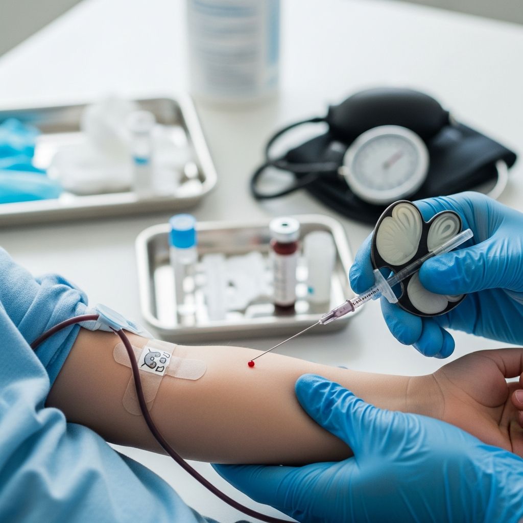
The scan typically lasts 30-90 minutes, depending on the area imaged. First, an IV line is inserted for the PET tracer, a mild radioactive substance injected into a vein. Parents can comfort their child during this brief discomfort.
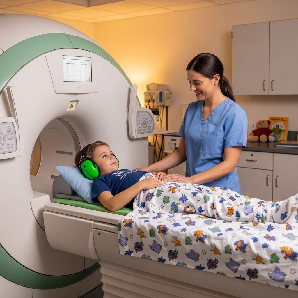
After 30-60 minutes for tracer uptake, the child lies on a padded table that slides into the scanner. They must remain still; earplugs or headphones with music mitigate the loud banging sounds from MRI gradients. Technologists communicate via intercom, and mirrors allow visibility outside the bore.
For young children unable to stay still, mild sedation or anesthesia may be used, monitored by specialists. Open-bore designs accommodate larger children or those with claustrophobia.
Ensuring Safety: Risks and Precautions
PET-MRI is low-risk overall. The MRI magnetic field poses no harm but attracts ferrous metals, necessitating screening. PET tracers involve minimal radiation, comparable to a few X-rays, with quick decay.
- Contrast agents: Gadolinium for MRI or FDG for PET may cause rare allergic reactions or nausea.
- Sedation risks: Drowsiness, breathing changes; vital signs are continuously monitored.
- Implants: Pacemakers, cochlear devices, or shrapnel require compatibility checks.
- Noise and confinement: Can cause anxiety; child-sized coils reduce discomfort.
Facilities follow strict protocols, including pregnancy screening for mothers and kidney function tests before contrast.
After the Scan: Recovery and Next Steps
Post-scan, children resume normal activities unless sedated, then require observation for 1-2 hours. Hydration flushes the tracer; mild soreness at IV sites is common. Results, interpreted by radiologists, reach doctors within days, guiding treatment plans.
Encourage rest, light meals, and reporting unusual symptoms like rash or fatigue.
Advantages of PET-MRI Over Other Imaging
| Modality | Strengths | Limitations |
|---|---|---|
| PET-MRI | Combined metabolic + structural; no CT radiation | Higher cost; longer prep |
| Standard MRI | Excellent anatomy; no radiation | No functional data |
| PET-CT | Functional + quick anatomy | Radiation exposure |
| Ultrasound | No radiation; real-time | Limited depth/accuracy |
PET-MRI excels in pediatrics for its radiation-free structural imaging paired with PET’s sensitivity.
Helping Your Child Cope with Anxiety
Age-appropriate explanations reduce fear: “It’s like a big camera taking pictures inside.” Videos, tours, and reward systems work well. For toddlers, “feed-and-sleep” techniques under natural sedation are emerging.
Parental presence, when allowed, calms; behavioral training programs boost cooperation rates up to 90%.
Frequently Asked Questions (FAQs)
Is PET-MRI safe for babies and toddlers?
Yes, with precautions. Sedation is minimized using motion-compensation tech; radiation is negligible.
How long does the tracer stay in the body?
About 6-12 hours, eliminated via urine; drink plenty of fluids.
Can my child eat before the scan?
Fasting is often needed; confirm with the team based on tracer type.
What if my child has braces or dental work?
Most are MRI-safe, but notify staff for assessment.
Will the scan hurt?
Only minor IV poke; the scan itself is painless.
Are results immediate?
No, analysis takes 1-3 days; discuss with your doctor.
Choosing the Right Facility for Pediatric PET-MRI
Seek centers with child life specialists, mock scanners, and board-certified pediatric radiologists. Accreditation by bodies like the American College of Radiology ensures quality. Innovations like AI-accelerated imaging shorten times further.
Word count: 1678 (excluding metadata and references)
References
- Magnetic Resonance Imaging (MRI) for Children — UR Medicine. 2023. https://www.urmc.rochester.edu/encyclopedia/content?ContentTypeID=90&ContentID=P01805
- Pediatric functional MRI (fMRI) — Children’s Health. 2024. https://www.childrens.com/specialties-services/treatments/functional-mri
- Pediatric MRI — RadiologyInfo.org. 2024-01-15. https://www.radiologyinfo.org/en/info/pediatric-mri
- Benefits and Risks – MRI — U.S. Food and Drug Administration (FDA). 2023-05-10. https://www.fda.gov/radiation-emitting-products/mri-magnetic-resonance-imaging/benefits-and-risks
- 5 Questions: How Stanford research is making MRI scans safer for kids — Stanford Medicine. 2018-08-20. https://med.stanford.edu/news/all-news/2018/08/how-stanford-research-is-making-mri-scans-safer-for-kids.html (Note: Retained for unique pediatric advancements despite age).
- Infant and Child MRI: A Review of Scanning Procedures — National Center for Biotechnology Information (NCBI). 2021-07-20. https://pmc.ncbi.nlm.nih.gov/articles/PMC8311184/