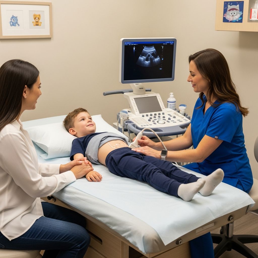
When a doctor recommends a pelvic ultrasound for a child or teen, many parents feel anxious and unsure of what the test involves. This guide explains, in simple terms, what a pelvic ultrasound is, why it is used, how to prepare, and what your child is likely to experience from start to finish.
Understanding Pelvic Ultrasound
A pelvic ultrasound is an imaging test that uses high-frequency sound waves to create pictures of organs and structures in the lower abdomen and pelvis, such as the uterus, ovaries, bladder, and surrounding tissues. The test does not use radiation and is considered very safe for infants, children, and adolescents.
How the Technology Works
During the exam, a small handheld device called a transducer sends sound waves into the body and listens for their echoes. A computer converts these echoes into real-time images on a screen.
- The sound waves are too high-pitched to be heard.
- A water-based gel is applied to the skin to help the sound waves travel between the transducer and the body.
- The technologist moves the transducer gently over the area being examined.
Why Ultrasound Is Often the First Choice
- No radiation: Unlike X-rays or CT scans, ultrasound uses sound, not ionizing radiation.
- Real-time imaging: It shows movement, such as blood flow and organ motion, as it happens.
- Noninvasive and painless: Most children feel only mild pressure from the transducer or the need to urinate because of a full bladder.
- Very useful in pediatrics: Ultrasound is considered the first-line imaging method for many conditions affecting the female pelvis in children and teens.
Why a Child Might Need a Pelvic Ultrasound
Doctors order pelvic ultrasounds for many reasons, usually to better understand the cause of symptoms or to follow up on abnormal exam findings. In pediatric and adolescent patients, pelvic ultrasound helps evaluate the uterus, ovaries, bladder, and sometimes nearby bowel and blood vessels.
Common Reasons in Children and Teens
- Pelvic or lower abdominal pain
- Pelvic mass or swelling felt during an exam
- Abnormal menstrual bleeding, such as very heavy periods or bleeding between cycles
- Very early puberty (precocious puberty) or lack of periods in a teen girl (primary amenorrhea)
- Urinary problems, such as frequent infections or difficulty urinating
- Evaluation after trauma or suspected infection in the pelvis
Pelvic ultrasound may also be used to monitor known conditions, such as ovarian cysts or certain congenital (present from birth) differences in the reproductive organs.
Types of Pelvic Ultrasound Approaches
The same ultrasound principles are used in every pelvic scan, but the way the transducer is placed can differ. The choice depends on the child’s age, developmental stage, and the clinical question.
| Approach | How It Is Done | Who Typically Gets It |
|---|---|---|
| Transabdominal | Transducer is moved over the lower abdomen on top of the skin with gel. | Infants, children, teens; first choice for most pediatric pelvic exams. |
| Transvaginal | Thin, covered transducer is gently placed into the vagina to obtain close-up views. | Sexually active adolescents and adults, when more detail is needed; usually not used in young children. |
Transabdominal Pelvic Ultrasound
For most children, a transabdominal scan is sufficient. The child lies on their back while the technologist moves the transducer over the lower belly.
- A full bladder is usually required to improve the view.
- The fluid in the bladder helps move gas-filled bowel out of the way and acts as a window so the pelvic organs can be seen clearly.
- This approach is completely external and does not enter the body.
Transvaginal Pelvic Ultrasound
In some older adolescents, especially those who are sexually active, a transvaginal ultrasound may be suggested when transabdominal views do not answer the clinical question.
- The probe is long and thin, covered with a sheath, and coated with gel.
- It is inserted a short distance into the vagina and gently moved to capture detailed images.
- Only done with the patient’s informed consent and explanation of what to expect.
Preparing Your Child for the Exam
Preparation is simple but important, particularly for transabdominal pelvic ultrasound. Instructions may vary slightly by hospital, but some common steps are listed below.
Bladder Preparation
- Full bladder for transabdominal scans: Many centers ask children to drink water before the appointment and avoid urinating so the bladder is full.
- If a child cannot reliably hold urine, the bladder may be filled using a temporary catheter in some cases.
- For transvaginal scans, the patient is usually asked to empty the bladder first.
Clothing and Personal Items
- Dress your child in comfortable, loose-fitting clothes.
- They may need to change into a hospital gown and remove jewelry or metal near the area being scanned.
- Conceal diapers or underwear may stay on or off depending on age and the specific exam; staff will explain what is needed.
Helping Your Child Feel Ready
Explaining the test in age-appropriate language can greatly reduce anxiety.
- Describe the ultrasound as a special camera that uses sound to take pictures inside the body.
- Reassure your child that they will be able to lie next to you or hold your hand in most settings.
- Bring comfort items such as:
- A favorite stuffed animal or blanket
- Books, a tablet, or music with headphones
- A snack for after the test if allowed
What Happens During the Pelvic Ultrasound
The exact routine varies by hospital, but most children have a similar experience.
Step-by-Step Overview
- Check-in and questionnaire: Families complete basic health and safety questions and confirm any preparation instructions.
- Changing clothes: If needed, your child changes into a gown and removes items that could interfere with the exam.
- Positioning on the table: Your child lies on their back on a padded exam table.[10]
- Applying the gel: Warm gel is placed on the lower abdomen (or on the transvaginal transducer for older adolescents).[10]
- Scanning: The technologist moves the transducer gently to obtain images from different angles.
- Occasional instructions: Your child may be asked to stay very still, briefly hold their breath, or shift position.
- Completion: The gel is wiped off and your child can get dressed.
The exam typically takes 20–40 minutes, depending on the child’s cooperation, the need for additional images, and whether more than one technique is used.
What Your Child Might Feel
- Cool or slightly warm gel on the skin
- Mild pressure as the transducer is pressed against the abdomen
- Strong urge to urinate from having a full bladder
- For transvaginal scans in older adolescents: possible brief discomfort or pressure, but usually not pain
If your child experiences pain, they should tell the technologist so the pressure or positioning can be adjusted.
What the Ultrasound Can Show
The person performing the scan focuses on capturing high-quality images. A radiologist or pediatric imaging specialist later analyzes these pictures in detail.
Structures Commonly Evaluated
- Bladder: Size, thickness of the wall, how well it empties, and any unusual masses or stones.
- Uterus: Size, shape, and the appearance of the inner lining, especially in adolescent girls.
- Ovaries: Size, number and type of follicles or cysts, and blood flow.
- Surrounding tissues: Fluid collections, masses, or signs of inflammation or torsion.
Possible Conditions Investigated
Pelvic ultrasound does not by itself provide a diagnosis in every case, but it gives crucial clues that help your child’s doctor. It can help evaluate for:
- Ovarian cysts or tumors
- Uterine or vaginal anomalies present from birth
- Pelvic inflammatory disease or abscesses in adolescents
- Ovarian torsion (twisting of the ovary that reduces blood flow)
- Causes of abnormal bleeding or pelvic pain
In emergency settings, ultrasound can also support rapid assessment for conditions such as ectopic pregnancy in teens.
Safety, Risks, and Comfort
Ultrasound is considered one of the safest imaging techniques available, especially for children.
Safety Profile
- No ionizing radiation: This is especially important in growing children, where limiting radiation exposure is a priority.
- Decades of experience: Ultrasound has been used in pediatrics and obstetrics for many years with a strong safety record when performed properly.
- Noninvasive: Most exams are performed entirely on the skin surface and do not break the skin or enter the body.
Potential Discomforts
- Temporary pressure on the abdomen
- Discomfort from holding urine in a full bladder
- Mild pressure or brief discomfort during transvaginal scans in appropriate older adolescents
Serious complications are extremely rare when ultrasound is used by trained professionals and appropriate medical guidelines are followed.
After the Ultrasound: Results and Next Steps
Most children can immediately go back to their usual activities after the exam.
Right After the Test
- Your child can use the restroom as soon as the images are complete.
- They may eat and drink normally unless another test with restrictions is scheduled.
- No recovery time or special monitoring is usually needed.
Getting the Results
- A radiologist or pediatric imaging specialist reviews and interprets the images.
- A report is sent to the ordering doctor, who will discuss the findings with you.
- Depending on the results, your child’s doctor may:
- Reassure you that the scan was normal
- Recommend follow-up imaging at a later date
- Order blood tests or other studies
- Refer you to a pediatric subspecialist, such as a pediatric gynecologist or urologist
Practical Tips for Parents and Caregivers
Small steps can make the visit smoother and less stressful for your child.
- Arrive early: Allow time for check-in, bathroom visits, and explaining the procedure calmly.
- Bring comfort items: Favorite toys or electronics can help distract and soothe.
- Stay calm and honest: Children often mirror their caregiver’s emotions; simple, truthful explanations help build trust.
- Ask questions: Technologists and doctors are used to explaining tests to families—use their expertise.
- Clarify follow-up: Before leaving, be sure you know how and when you will receive results.
Frequently Asked Questions (FAQs)
Is a pelvic ultrasound painful for children?
A pelvic ultrasound is usually not painful. Your child may feel mild pressure from the transducer and the discomfort of a full bladder, but the test itself is generally well tolerated.
How long does a pediatric pelvic ultrasound take?
Most pelvic ultrasounds take about 20–40 minutes from the time your child lies on the table until the images are complete, depending on cooperation and the complexity of the case.
Are there any risks or side effects?
Ultrasound has an excellent safety record and does not use radiation. Side effects are rare and usually limited to temporary discomfort from pressure or a full bladder.
Will my child be sedated?
Sedation is not typically needed for pelvic ultrasound. In rare cases with very young or extremely anxious children, other calming strategies may be discussed, but most children manage well with reassurance and distraction.
When will we get the results?
A radiologist reviews the images and sends a report to the doctor who ordered the exam. That doctor will usually discuss results with you within a short time frame, which can vary by hospital and urgency.
References
- Sonography Pediatric Gynecology Assessment, Protocols, and Interpretation — Mellerio G, et al., StatPearls Publishing (NCBI Bookshelf). 2023-07-24. https://www.ncbi.nlm.nih.gov/books/NBK570595/
- Pelvic ultrasound — Children’s Healthcare of Atlanta. 2021-06-01. https://www.choa.org/-/media/Files/Childrens/teaching-sheets/pelvic-ultrasound.pdf
- Pelvis Ultrasound — RadiologyInfo.org (Radiological Society of North America / American College of Radiology). 2022-09-01. https://www.radiologyinfo.org/en/info/pelvus
- Ultrasound Scan — Nationwide Children’s Hospital. 2020-10-15. https://www.nationwidechildrens.org/family-resources-education/health-wellness-and-safety-resources/helping-hands/ultrasound-scan
- Pelvic Ultrasound — Stanford Medicine Children’s Health. 2022-03-10. https://www.stanfordchildrens.org/en/topic/default?id=pelvic-ultrasound-92-P07784
- Pediatric Ultrasound — Children’s Hospital of Philadelphia. 2021-09-01. https://www.chop.edu/treatments/ultrasound
- Ultrasound Examinations & Preparations — Boston Children’s Hospital. 2019-11-12. https://www.childrenshospital.org/treatments/ultrasound