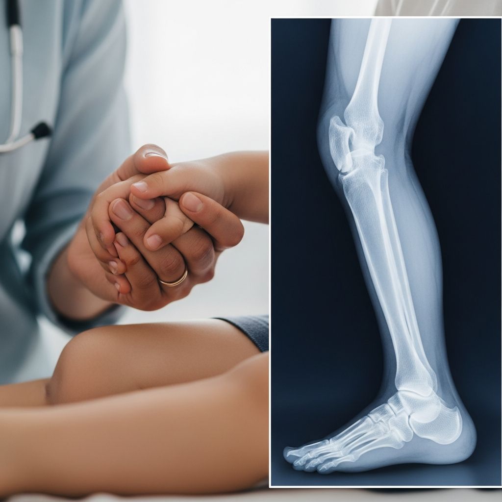
Lower leg X-rays are a cornerstone diagnostic tool in pediatric medicine, helping clinicians assess injuries, growth issues, and bone health in children. These imaging tests use low-dose radiation to create clear pictures of the tibia, fibula, knee, ankle, and foot structures. Parents often encounter this procedure after playground falls, sports mishaps, or concerns about limping.
Why Doctors Recommend Lower Leg X-Rays for Kids
Pediatricians order lower leg X-rays primarily to investigate suspected fractures from trauma, which are common in active children. These images reveal breaks in the tibia (shinbone) or fibula (smaller leg bone), as well as joint issues like effusions or dislocations. Beyond acute injuries, X-rays detect chronic conditions such as stress fractures from overuse in young athletes, infections like osteomyelitis, or alignment problems contributing to gait abnormalities.
In growing kids, these scans measure bone length discrepancies between legs, which can arise from congenital differences or post-injury growth plate disruptions. Early detection allows interventions like braces or surgery to prevent scoliosis or hip strain. Arthritis, though rarer in children, tumors, osteoporosis signs, or deformities also warrant imaging.
Anatomy Highlighted in Children’s Lower Leg X-Rays
The lower leg comprises the tibia and fibula, extending from knee to ankle. X-rays capture the knee joint (femur-tibia meeting), ankle mortise (tibia-fibula-talus alignment), and foot bones including tarsals, metatarsals, and phalanges. In kids, open growth plates appear as darker lines across bone ends, distinguishing pediatric from adult images.
Standard views include anteroposterior (AP, front-to-back), lateral (side), and oblique angles for comprehensive assessment. For knees, horizontal beam lateral views detect fluid levels signaling intra-articular fractures. Ankle imaging follows Ottawa rules to minimize unnecessary scans, focusing on tenderness points.
Preparing Your Child for a Lower Leg X-Ray
Preparation is straightforward and stress-free. No fasting or special diets are needed. Dress your child in loose clothing; remove shoes, socks, and metal jewelry like belts or braces from the leg area to avoid image artifacts. Explain the process simply: “It’s like a quick camera snap of your leg bones—no pain, just hold still like a statue.”
For anxious toddlers, bring a favorite toy or use distraction apps. Older kids benefit from knowing they’ll stand or lie briefly while the tech positions them. If your child has developmental needs, inform the radiology team ahead for accommodations.
- Arrive 15 minutes early for paperwork.
- Discuss pregnancy concerns if applicable (rare in kids).
- Pack snacks for post-scan comfort.
Step-by-Step: What Happens During the Procedure
The entire process lasts 10-20 minutes in a radiology suite, often at a children’s hospital or clinic. A certified radiologic technologist, trained in pediatric radiation safety, guides your child.
- Positioning: Child stands or lies on the X-ray table. Tech uses pillows or sandbags for stability, ensuring the knee points forward and ankle relaxes.
- Shielding: Lead aprons cover the torso, thyroid, and pelvis to block scatter radiation, prioritizing gonadal protection.
- Imaging: Tech retreats behind a screen and activates the machine for 2-4 views per leg. Hold breath for 1-2 seconds per shot to prevent blur.
- Repositioning: Quick adjustments for AP, lateral, and skyline (patella) views if needed.
For leg length checks, standing scans compare sides; blocks under the shorter leg simulate correction. Painful injuries? Tech accommodates gently.
Radiation Safety and Risks for Pediatric Patients
X-rays emit ionizing radiation, but pediatric doses are minimized using ALARA (As Low As Reasonably Achievable) principles. A single lower leg series equals background radiation from 2-3 days outdoors—safe for diagnostics. Digital detectors reduce exposure by 50-80% versus film.
Risks are negligible: no immediate side effects, lifetime cancer risk increase under 1 in 10,000 for one exam. Multiple scans? Discuss cumulative dose with your doctor. Pregnant guardians hold infants outside the room.
| Factor | Pediatric Benefit | Safety Measure |
|---|---|---|
| Dose | 0.001-0.01 mSv | Collimation limits beam |
| Views | 2-4 minimum | Evidence-based protocols |
| Shielding | Full body | Lead aprons mandatory |
Interpreting X-Ray Findings: What Radiologists Look For
A pediatric radiologist analyzes images systematically: alignment, bone cortex integrity, trabecular patterns, and soft tissues. Normal: Smooth cortices, aligned joints, symmetric growth plates.
Common abnormalities:
- Fractures: Tibial plateau (depressed cortex, lipohemarthrosis), patellar (skyline discontinuity), fibular head (high-energy marker).
- Ankle: Weber types A-C by fibula break level; syndesmosis stability.
- Foot: Metatarsal stress (periosteal reaction), calcaneal (Böhler’s angle loss), 5th MT base avulsion.
- Infection/Tumor: Bone erosion, periosteal lift.
Results reach your doctor within hours; follow-up MRI/CT if unclear.
After the X-Ray: Next Steps and Recovery
No recovery needed—resume activities immediately unless injury restricts. Discuss results promptly: fractures may need casting, alignment issues orthotics, or lengths surgery. Serial X-rays monitor healing/growth.
Watch for worsening pain/swelling; seek ER if needed.
Common Myths About Kids’ X-Rays Debunked
- Myth: X-rays hurt. Fact: Painless positioning only.
- Myth: Too risky for kids. Fact: Benefits outweigh minimal radiation.
- Myth: One view suffices. Fact: Multi-views essential for accuracy.
Frequently Asked Questions (FAQs)
Is lower leg X-ray safe for my 5-year-old?
Yes, with pediatric-optimized doses far below harmful levels.
How long do results take?
Preliminary reads in minutes; full report same day.
What if my child can’t stay still?
Techs use games, swaddling, or sedation rarely.
Can X-rays show soft tissue damage?
Limited; swelling/effusion clues, but MRI better.
Are follow-up X-rays needed?
Often yes, to track healing or growth.
Empowering Parents: When to Advocate for Imaging
Trust Ottawa ankle/knee rules: Image if specific tenderness/deformity. For subtle limps, push for scans to catch growth issues early. Partner with pediatric orthopedists for holistic care.
This guide equips you to navigate lower leg X-Rays confidently, ensuring your child’s bone health is addressed promptly and safely.
References
- Lower Limb X-Ray Interpretation — Mind The Bleep, Dr. Joe Kang. 2023. https://mindthebleep.com/lower-limb-x-ray-interpretation/
- Lower Extremity X-Ray Patient Guide — Sanitas Medical Group. 2022. https://www.mysanitas.com/sites/default/files/resources/lower_extremity_x-ray_patient_guide.pdf
- Foot X-Ray: Anatomy, Procedure & What to Expect — Cleveland Clinic. 2024-01-17. https://my.clevelandclinic.org/health/diagnostics/23500-foot-x-ray
- Lower Limb Alignment — Radiology Key. 2021. https://radiologykey.com/lower-limb-alignment/
- How Does a Leg Length X-Ray Work? — Dayton Children’s Hospital. 2023. https://childrensdayton.org/kidshealth/xray-leg-length/