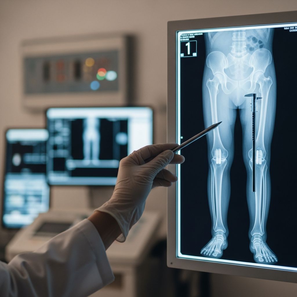
Leg length X-rays serve as a vital diagnostic tool in pediatric orthopedics, enabling healthcare providers to precisely evaluate differences in lower limb lengths. These imaging studies are particularly important for children, where uneven growth can lead to posture issues, pain, or gait abnormalities if not addressed early. By capturing detailed images of the femur, tibia, fibula, and surrounding structures, this exam helps quantify discrepancies and guide interventions ranging from observation to surgical options.
Why Leg Length Differences Occur in Kids
Uneven leg lengths, known as leg length discrepancy (LLD), affect up to 1 in 1000 children significantly enough to require monitoring. Causes include congenital conditions like hemihypertrophy, where one side of the body grows faster, or acquired issues such as fractures, infections, or conditions like Blount’s disease that alter bone development. In many cases, minor asymmetries are normal and self-correct with growth, but discrepancies over 1-2 cm can strain the hips, knees, spine, and cause scoliosis-like curvatures.
During rapid growth phases, such as puberty, these differences may become more pronounced. Early detection through X-rays allows orthopedists to predict final discrepancies at skeletal maturity using growth plate assessments called scanograms, helping families plan ahead.
The Science Behind Leg Length Measurement Techniques
Accurate LLD assessment combines clinical exams with imaging. Non-radiographic methods like block placement under the shorter leg provide initial estimates by leveling the pelvis, but they lack precision for bone-specific measurements. Radiographic approaches offer superior accuracy by visualizing the entire lower extremity.
- Teleoroentgenogram: A single full-length standing AP view from 180 cm distance, centered at the knee, minimizes magnification errors and captures weight-bearing alignment.
- CT Scanogram: Supine scout views measure femur and tibia separately with low radiation, ideal for serial monitoring in growing children.
- Computed Radiography (CR): Digital stitching of hip, knee, and ankle images with scaling markers ensures precise centimeter-level measurements.
Modern systems incorporate AI to automate femur/tibia lengths and detect pelvic tilt or axis deviations, streamlining radiologist workflows.
Preparing Your Child for the Exam
Preparation is straightforward to ensure clear images and minimize stress. Instruct your child to wear loose clothing and remove metal objects like belts or jewelry from the waist down. No fasting is required, and the exam typically lasts 5-15 minutes. Explain the process simply: “You’ll stand still like a superhero while the machine takes pictures of your legs to help the doctor.” Practice standing straight with knees forward.
For anxious kids, bring a favorite toy or use distraction techniques. If your child has mobility aids or special needs, inform the team in advance for accommodations like foam pads under feet.
Step-by-Step: What Happens During the Procedure
The exam occurs in a radiology suite with a technologist guiding every step. Your child stands on a platform with knees and ankles facing forward, patellae straight ahead. A ruler or scaling marker is positioned nearby for calibration. Multiple low-dose exposures—often three (hip, knee, ankle)—are taken and digitally combined into one composite image.
- Positioning: Patient stands erect or lies supine; blocks may elevate the shorter leg to assess pelvic leveling.
- Imaging: X-ray beam scans from pelvis to ankles; child holds still for seconds per shot.
- Review: Tech checks images on-site; repeats if needed for clarity.
- Completion: Child dresses and receives aftercare instructions.
Standing views best reflect functional LLD under weight-bearing, while supine positions reduce motion artifacts in young patients.
Radiation Safety and Risks in Pediatric Imaging
X-rays involve ionizing radiation, but pediatric doses for leg length studies are minimal—equivalent to 1-2 days of natural background exposure. Techniques like CR teleoroentgenograms deliver 1-2 mrad, far below harmful thresholds, making them suitable for repeated use in growth monitoring.
Risks are negligible when clinically justified, as benefits like preventing chronic pain or deformity outweigh exposure. The ALARA principle (As Low As Reasonably Achievable) guides protocols: collimation limits the beam, digital detectors enhance efficiency. No sedation is needed, avoiding additional risks.
| Method | Radiation Dose | Best Use Case |
|---|---|---|
| Teleoroentgenogram | Low (1-2 mrad) | Initial weight-bearing eval |
| CT Scanogram | Very low | Serial measurements |
| Stitched AP Views | Minimal | Routine clinical |
Interpreting the Results: What Doctors Look For
Radiologists measure from femoral head to ankle plafond, comparing sides. Discrepancies under 1 cm often need no intervention; 2+ cm may warrant epiphysiodesis (growth plate arrest) or lengthening surgery. Images also reveal angular deformities, joint pathologies, or growth plate status.
Follow-up X-rays track progression, especially near growth plate closure around ages 12-16 for girls and 14-18 for boys. AI-assisted analysis flags mechanical axis deviations for precise alignment correction.
Treatment Pathways After Diagnosis
Options depend on discrepancy size, growth remaining, and symptoms:
- Observation: For minor LLD (<2 cm), periodic scans suffice.
- Non-surgical: Shoe lifts correct functional inequality without surgery.
- Surgical: Temporary hemiepiphysiodesis halts growth on the longer leg; osteotomies or external fixators for severe cases.
Multidisciplinary teams including orthopedists, radiologists, and physical therapists optimize outcomes.
Common Concerns Addressed: Parental FAQ
Is the X-ray painful for my child?
No, it’s painless—just requires standing still. Young kids may find it briefly uncomfortable but not hurtful.
How accurate are these measurements?
Highly accurate (<0.5 cm error) with digital markers and standing protocols, superior to tape measures.
Can repeated X-rays harm growing bones?
Doses are safe for monitoring; radiation is far lower than a cross-country flight.
What if no discrepancy is found?
Images may still spot other issues like fractures or alignment problems explaining symptoms.
When should we worry about leg length?
If limping, hip/knee pain, or uneven shoulders/hips appear; consult a pediatric orthopedist.
Are there alternatives to X-rays?
Ultrasound for infants or EOS low-dose biplanar imaging, but X-rays remain gold standard for school-age kids.
Long-Term Outlook and Monitoring Growth
Most children with mild LLD achieve normal function with timely care. Regular check-ups predict adult leg lengths via Risser grading of iliac apophyses. Families should track shoe wear patterns and gait at home between visits.
Advances like 3D biplanar radiography promise even lower radiation and 3D modeling for complex cases, enhancing precision.
Empowering Parents: Next Steps Post-Exam
Results are typically available within days via your orthopedist. Discuss management plans openly, including lifestyle adjustments like balanced sports participation to avoid overuse injuries. Support groups for limb discrepancies offer emotional resources.
By understanding this exam, parents play a key role in their child’s musculoskeletal health, ensuring proactive steps toward even mobility and pain-free living.
References
- Methods for Assessing Leg Length Discrepancy — Sabharwal S, et al. National Center for Biotechnology Information (PMC). 2008-12-01. https://pmc.ncbi.nlm.nih.gov/articles/PMC2628227/
- The Radiological X-ray Exam: Leg Length Measurements — East Sussex Hospitals NHS Trust. 2022-01-01. https://www.esht.nhs.uk/wp-content/uploads/2022/01/0951.pdf
- Leg Length X-Ray Exam — Nemours KidsHealth. Recent access 2026. https://kidshealth.org/en/parents/xray-leg-length.html
- Leg Length Discrepancy AI Use Case — American College of Radiology. Recent access 2026. https://www.acr.org/Data-Science-and-Informatics/AI-in-Your-Practice/AI-Use-Cases/Use-Cases/Leg-Length-Discrepancy
- Assessment of Lower Limb Length and Alignment by Biplanar — American Journal of Roentgenology. 2013-01-01. https://ajronline.org/doi/10.2214/AJR.13.10782
- Leg Length Differences — Texas Children’s Hospital. Recent access 2026. https://www.texaschildrens.org/sites/default/files/Leg-Length-Differences.pdf